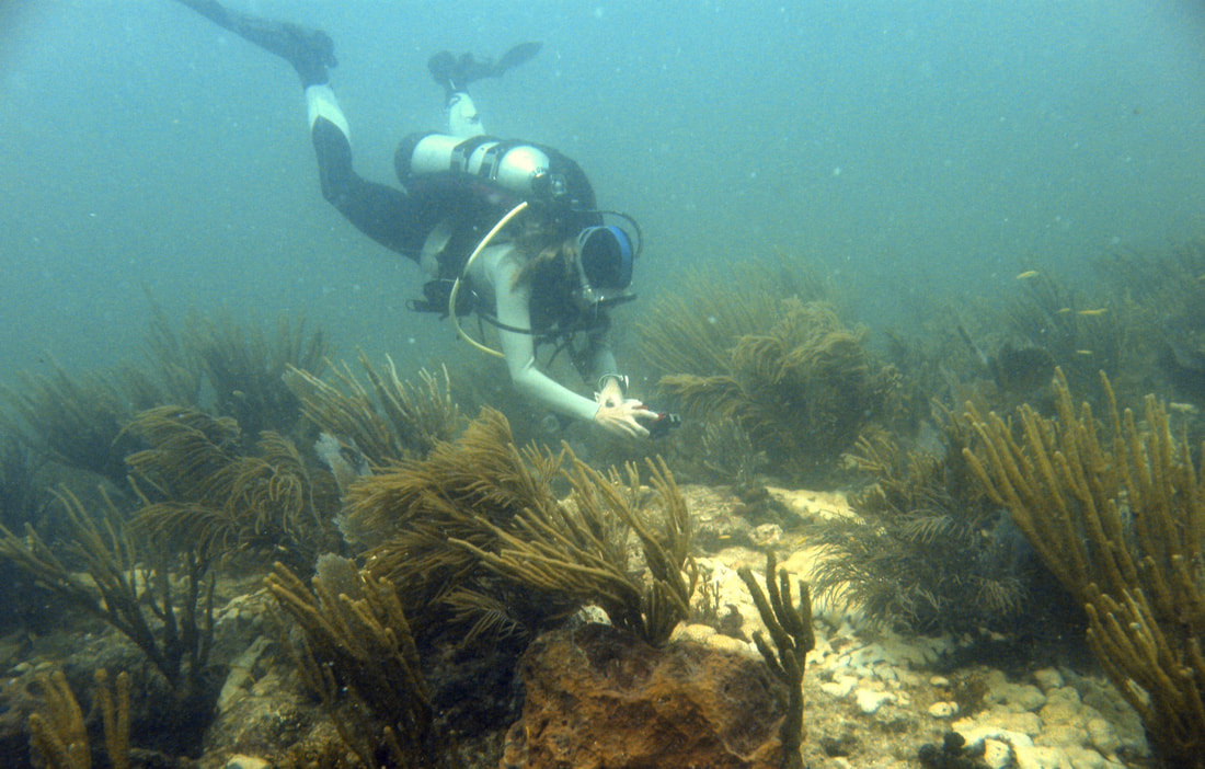
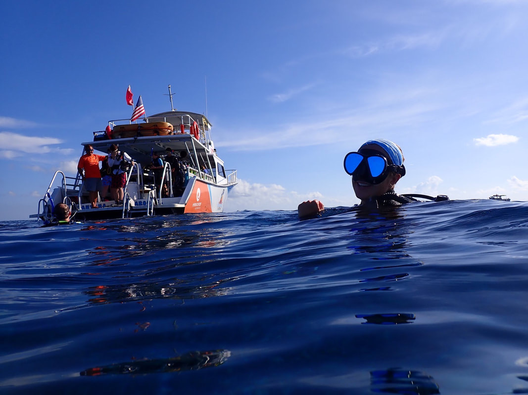
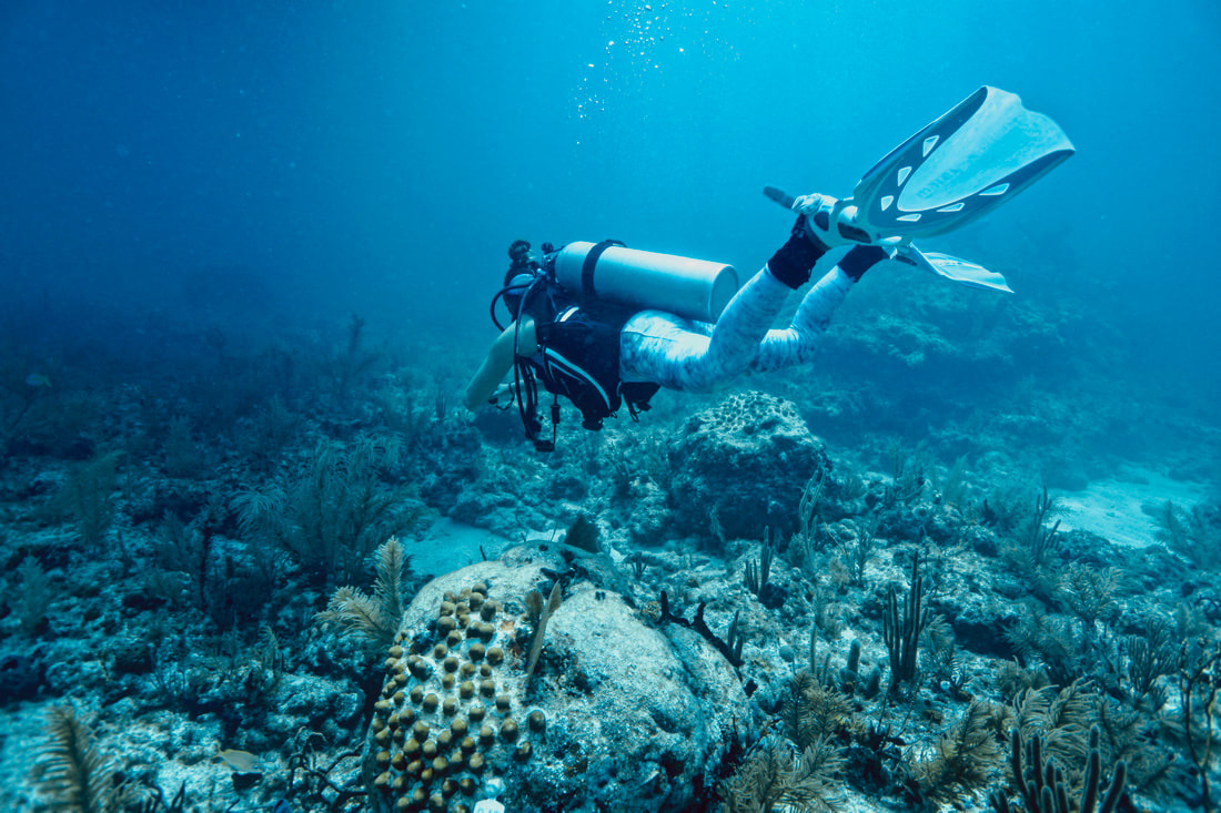
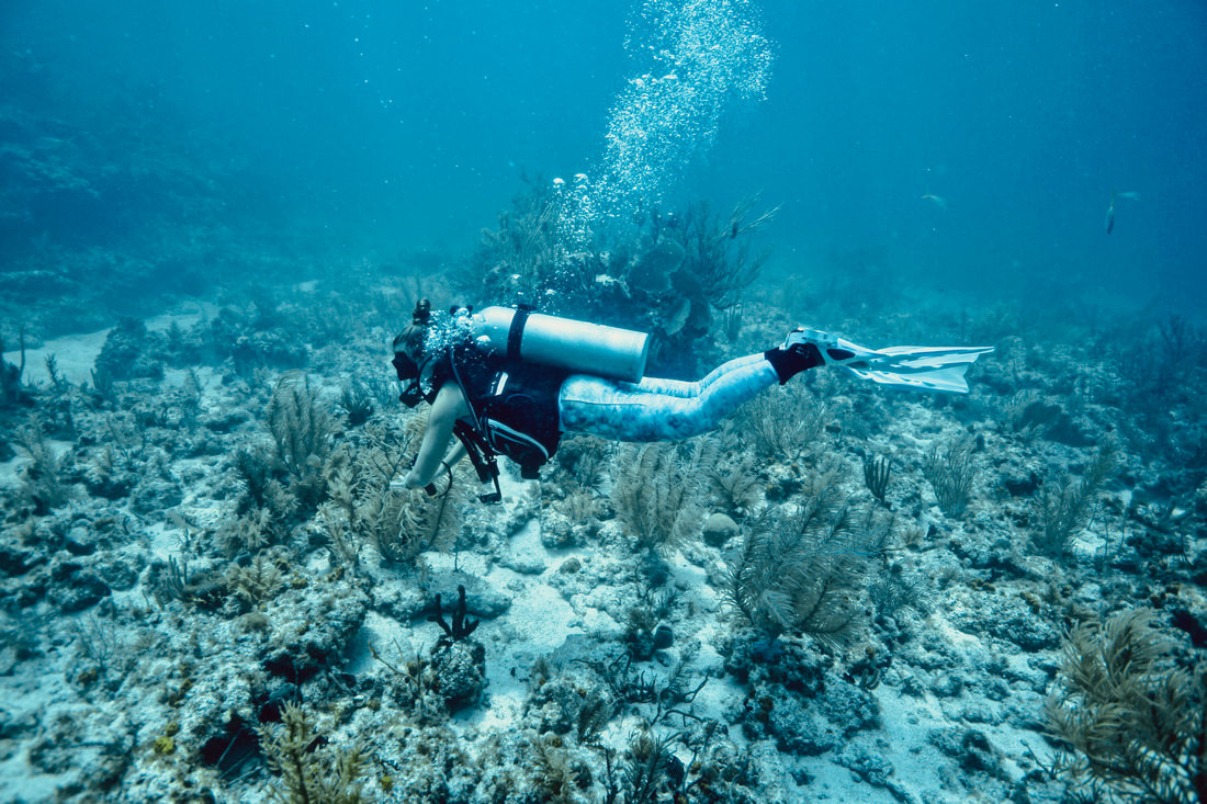
Ever since I became open-water certified in February 2018 in Hawai’i, I’ve been fascinated with scuba diving and underwater photography. I started with a GoPro 4 and now use a GoPro HERO 7.
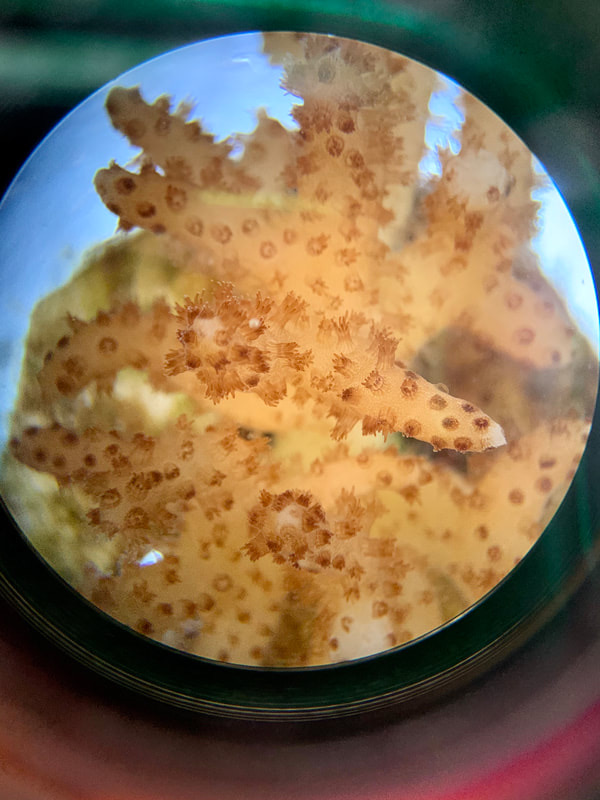
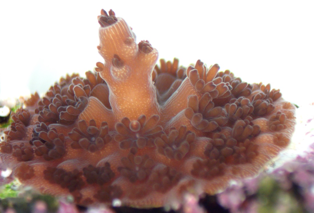
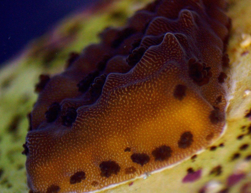
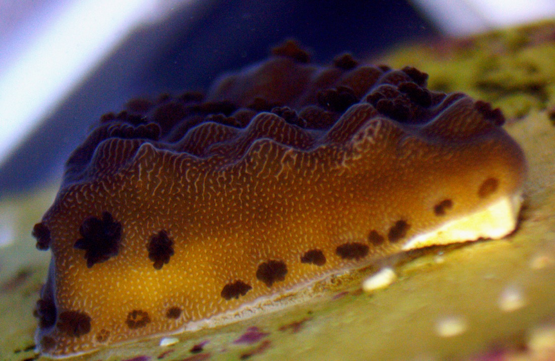
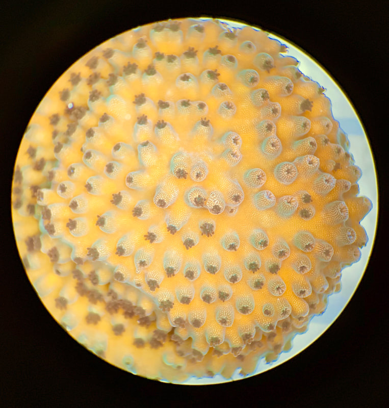
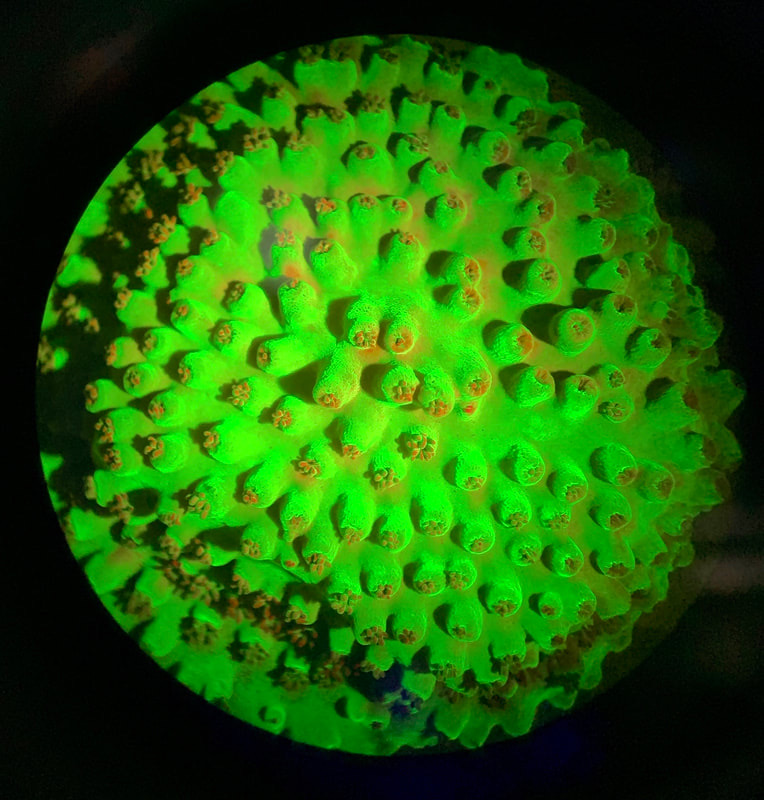
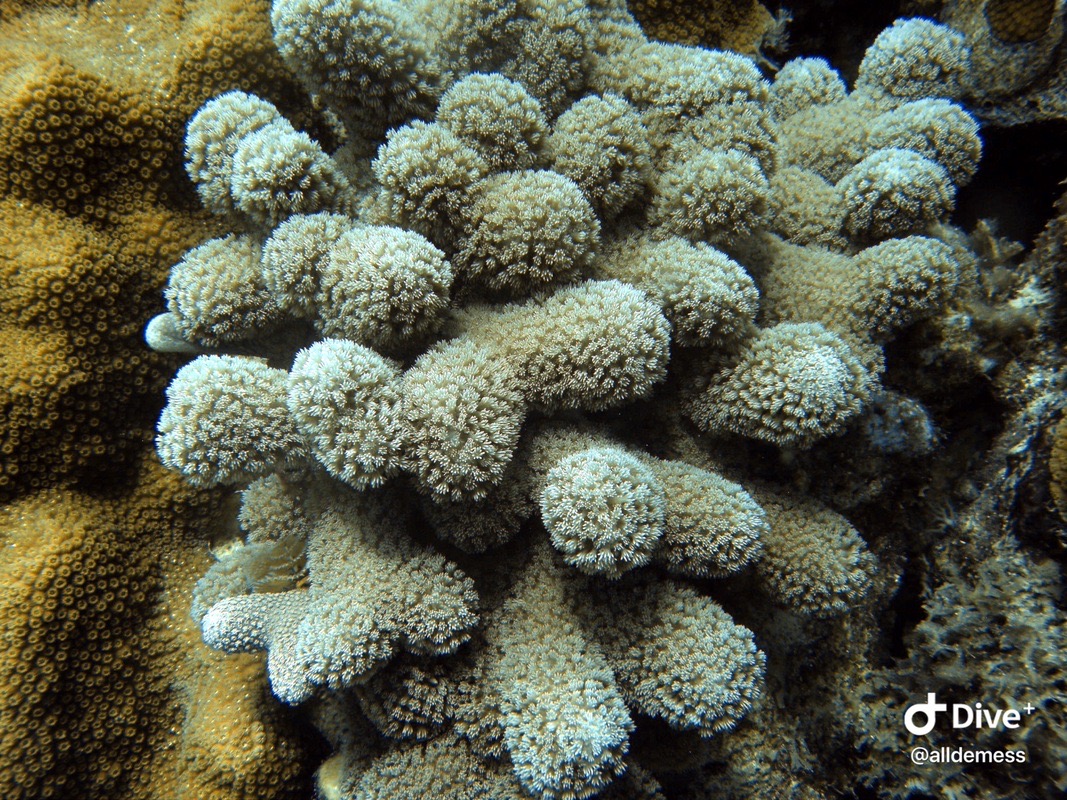
Pocillopora damicornis (a Pacific coral)
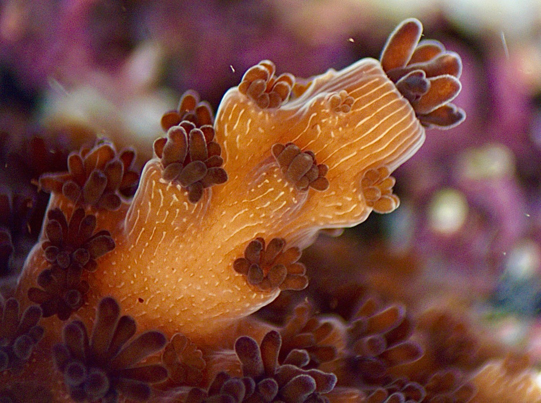
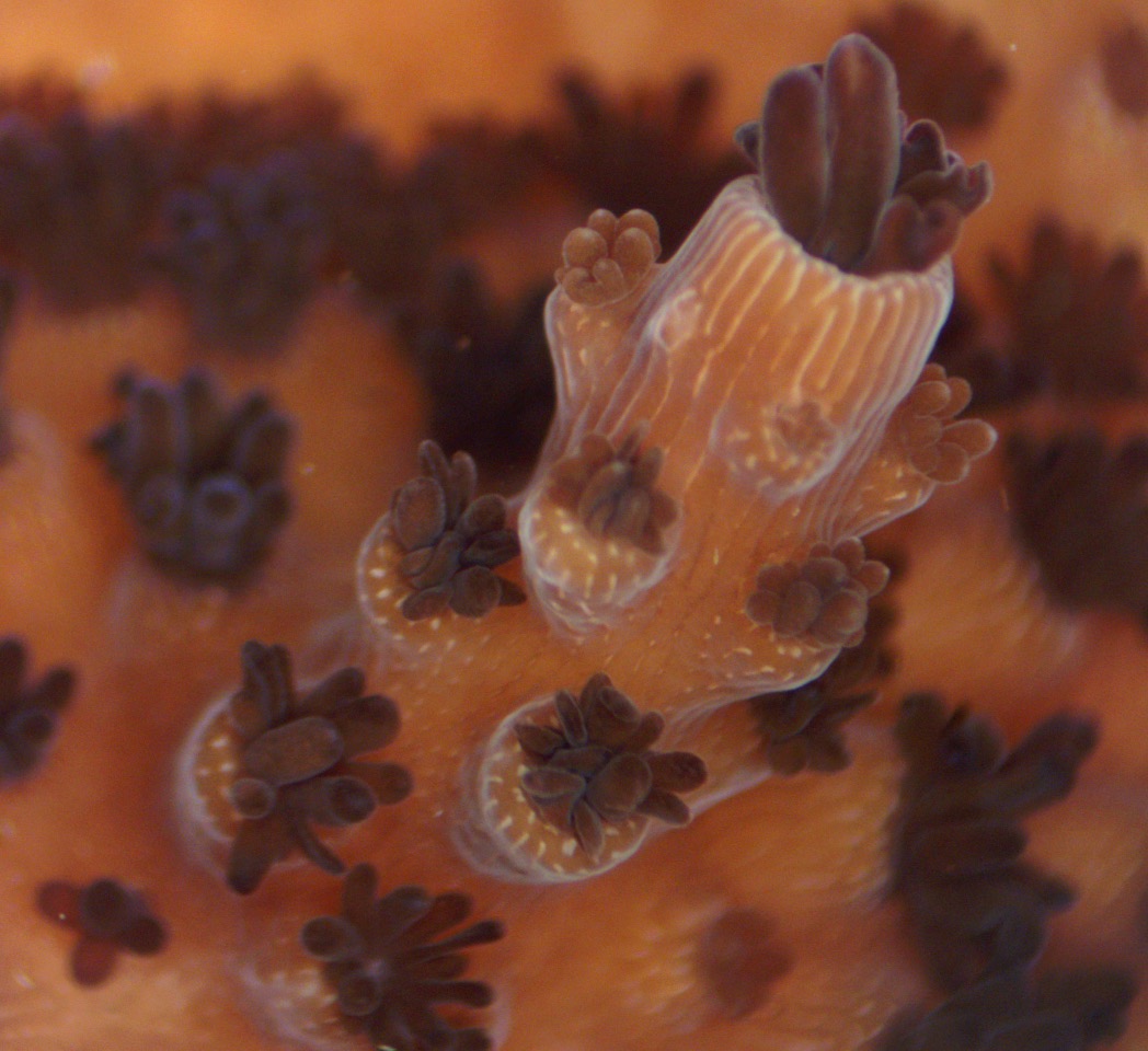
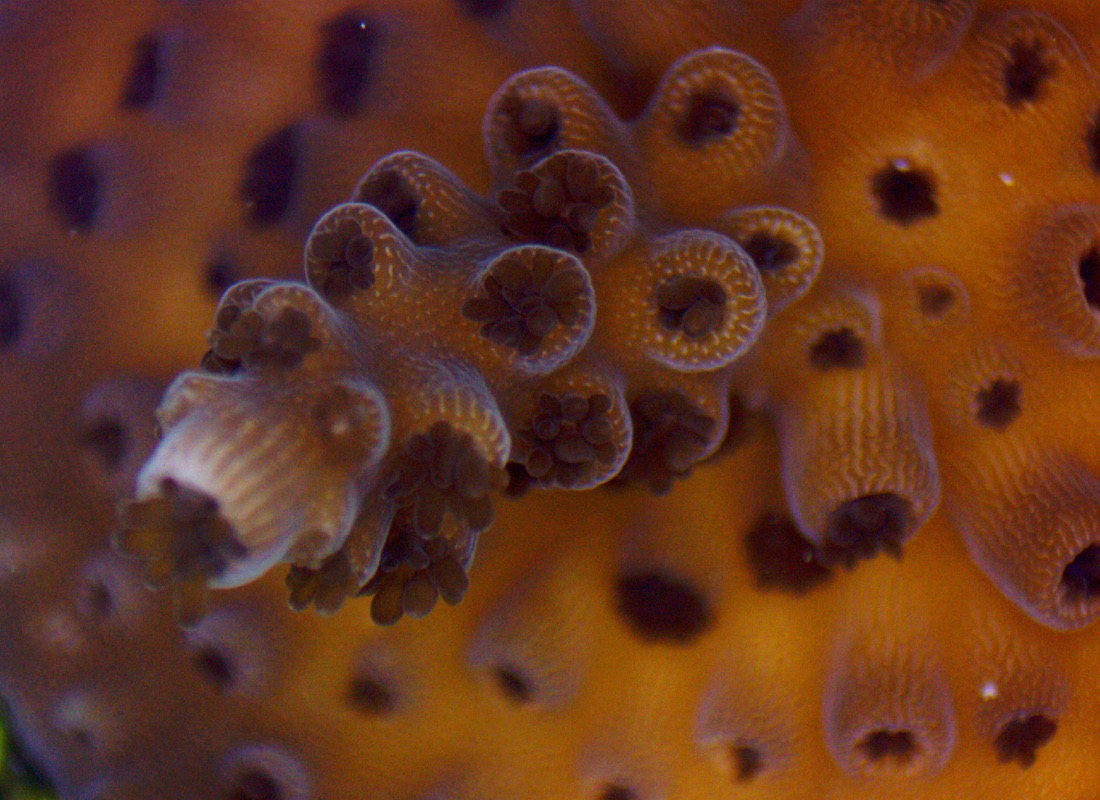
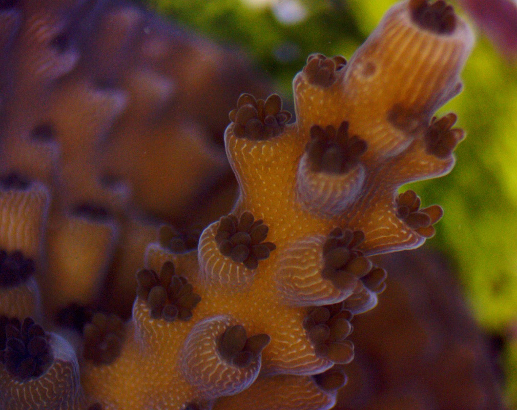
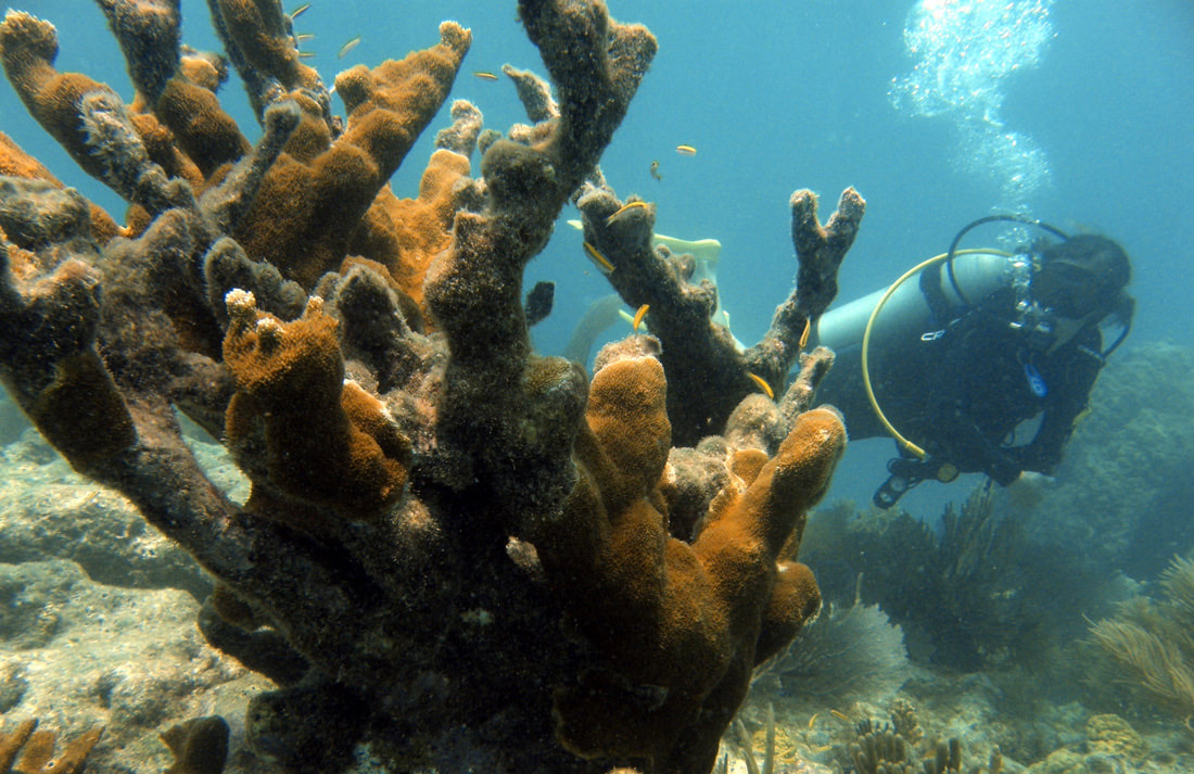
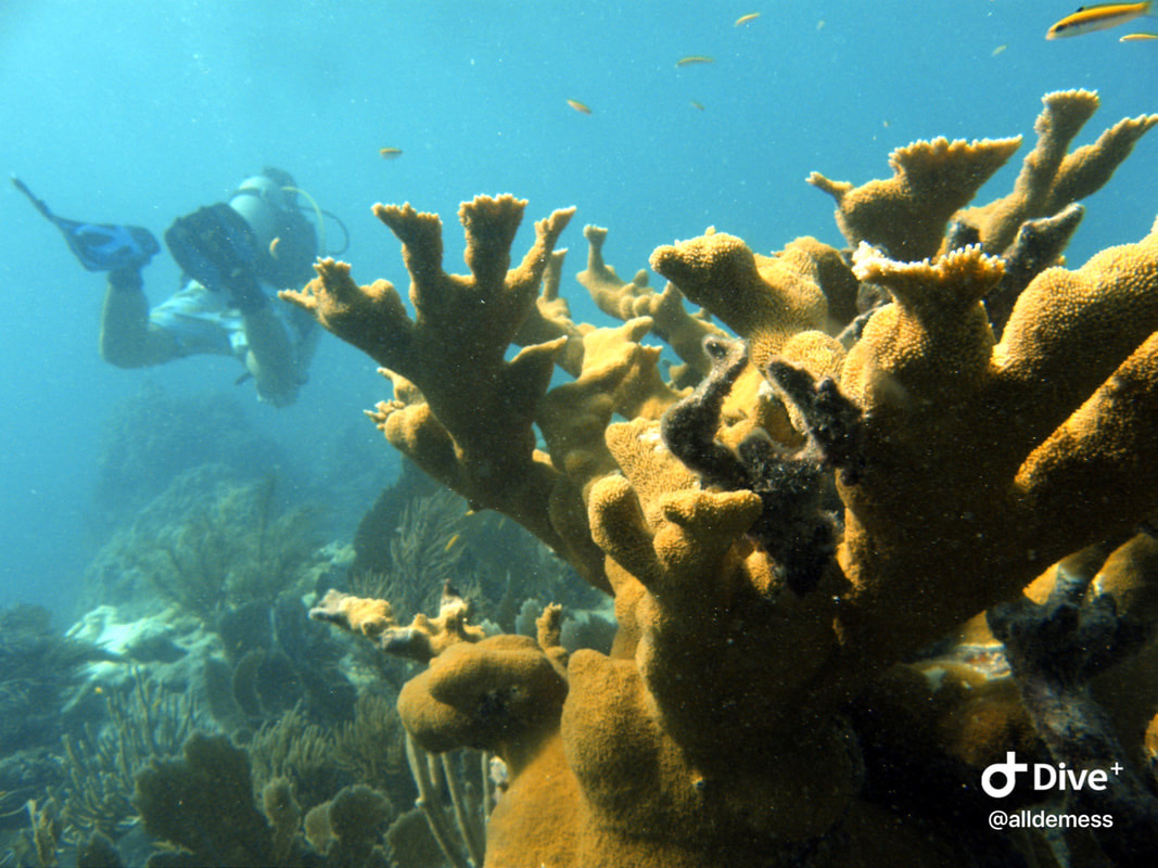
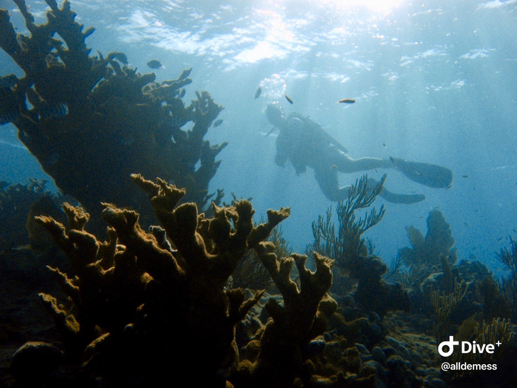
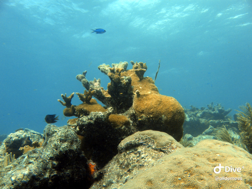
Acropora cervicornis (staghorn coral)
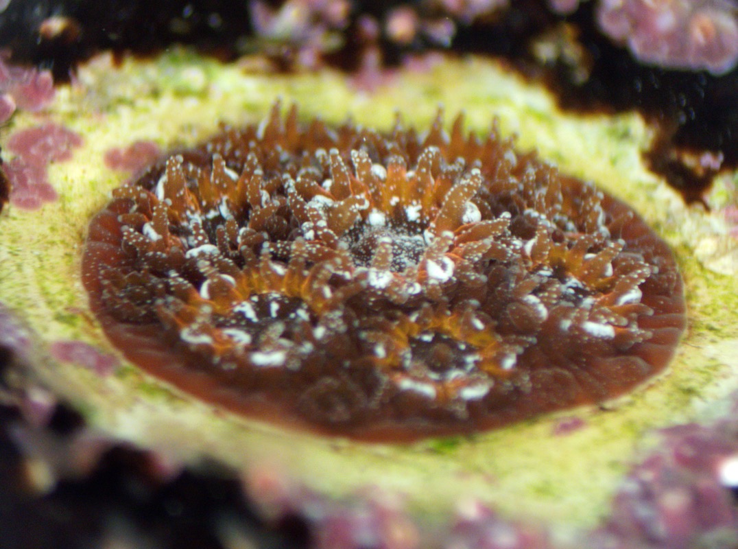
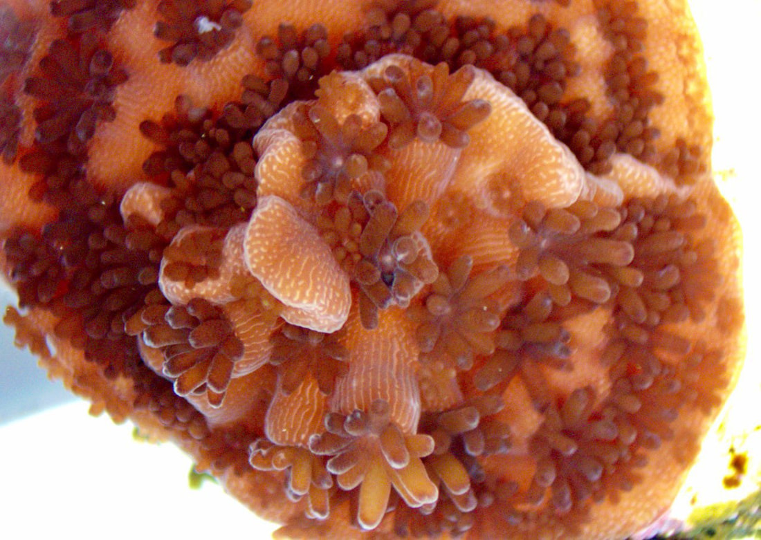
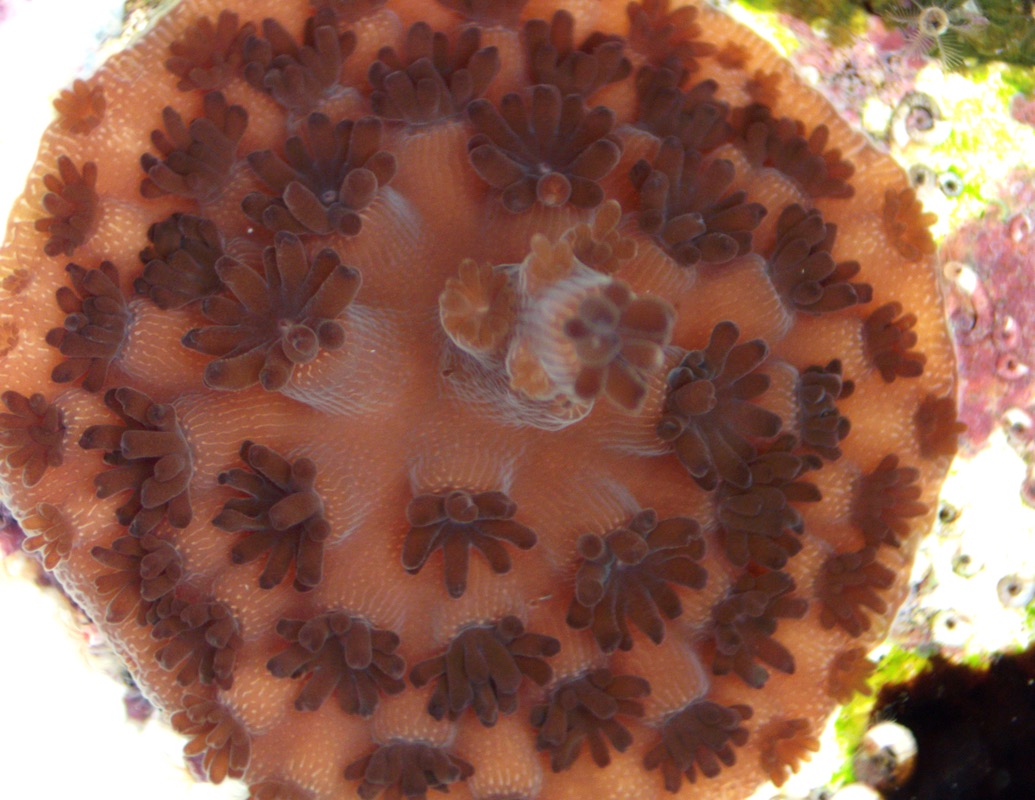
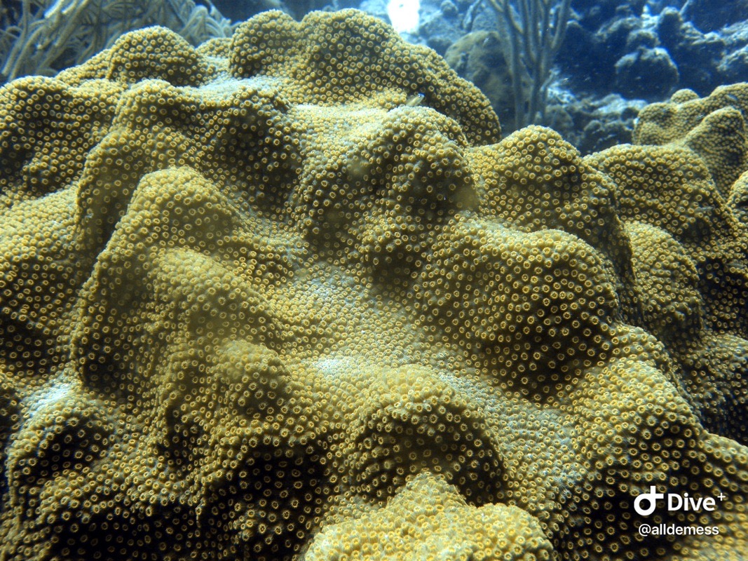
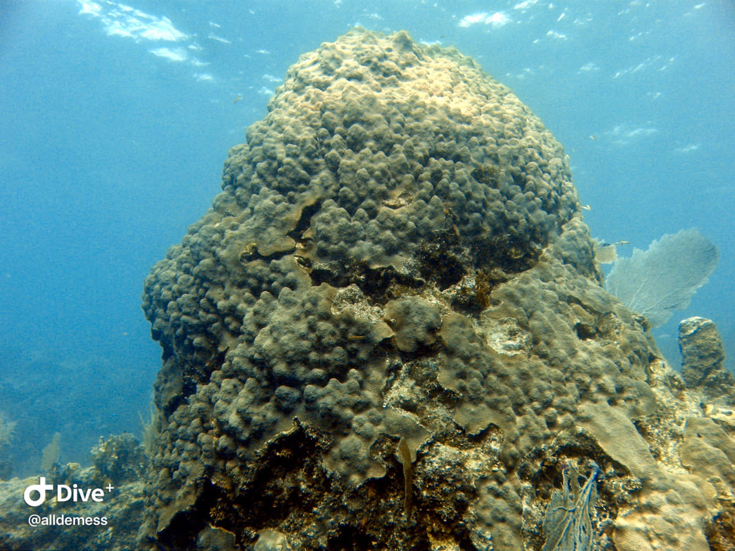
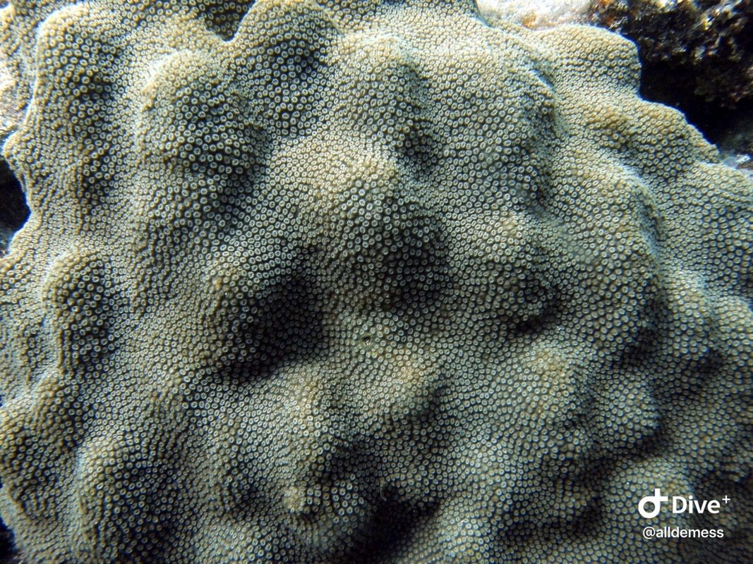
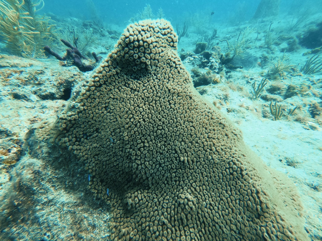
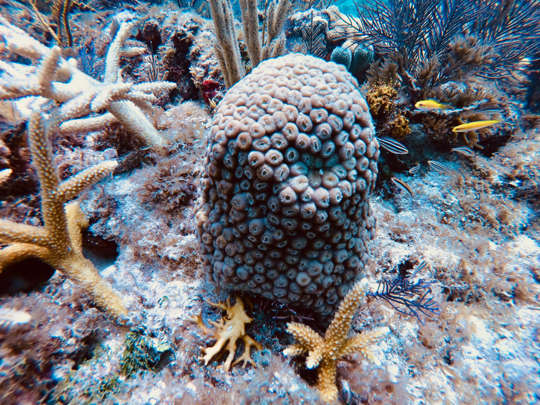
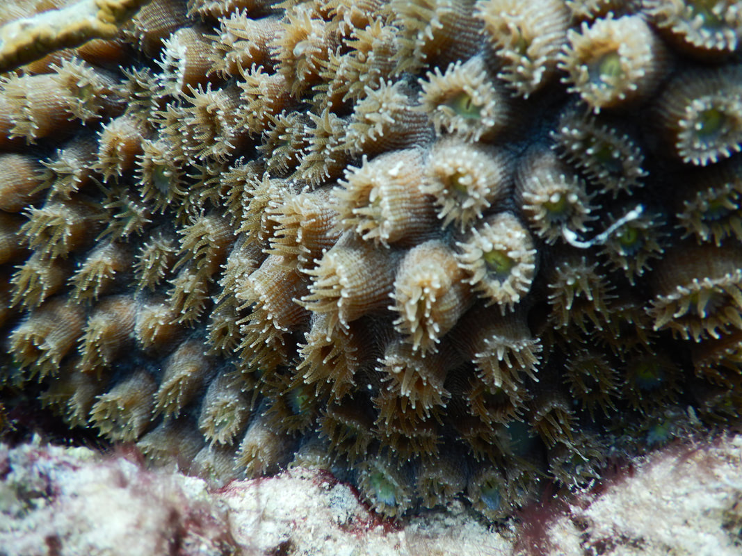
Orbicella faveolata (Mountainous star coral)
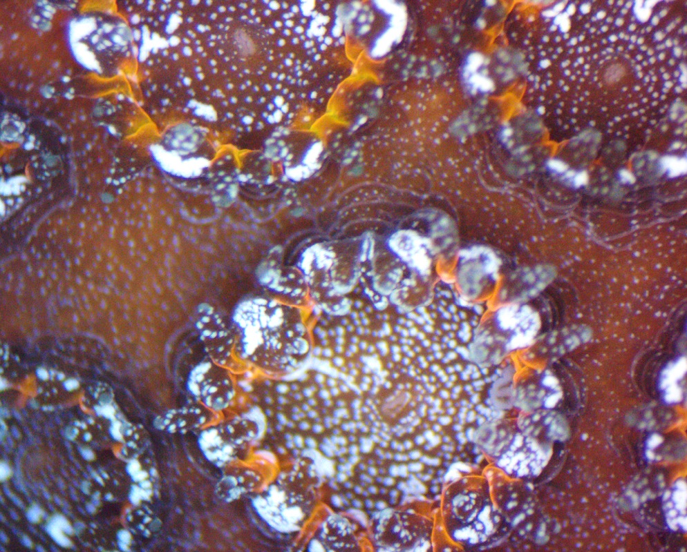
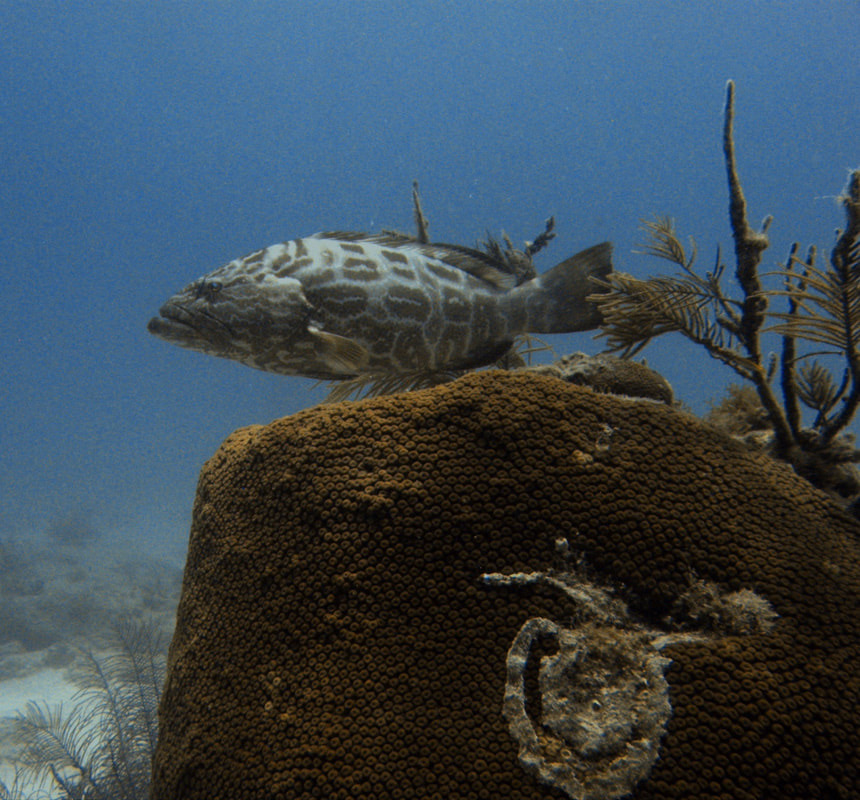
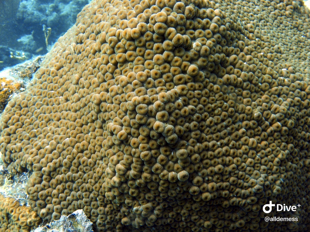
Orbicella faveolata (Mountainous star coral)
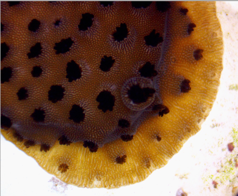
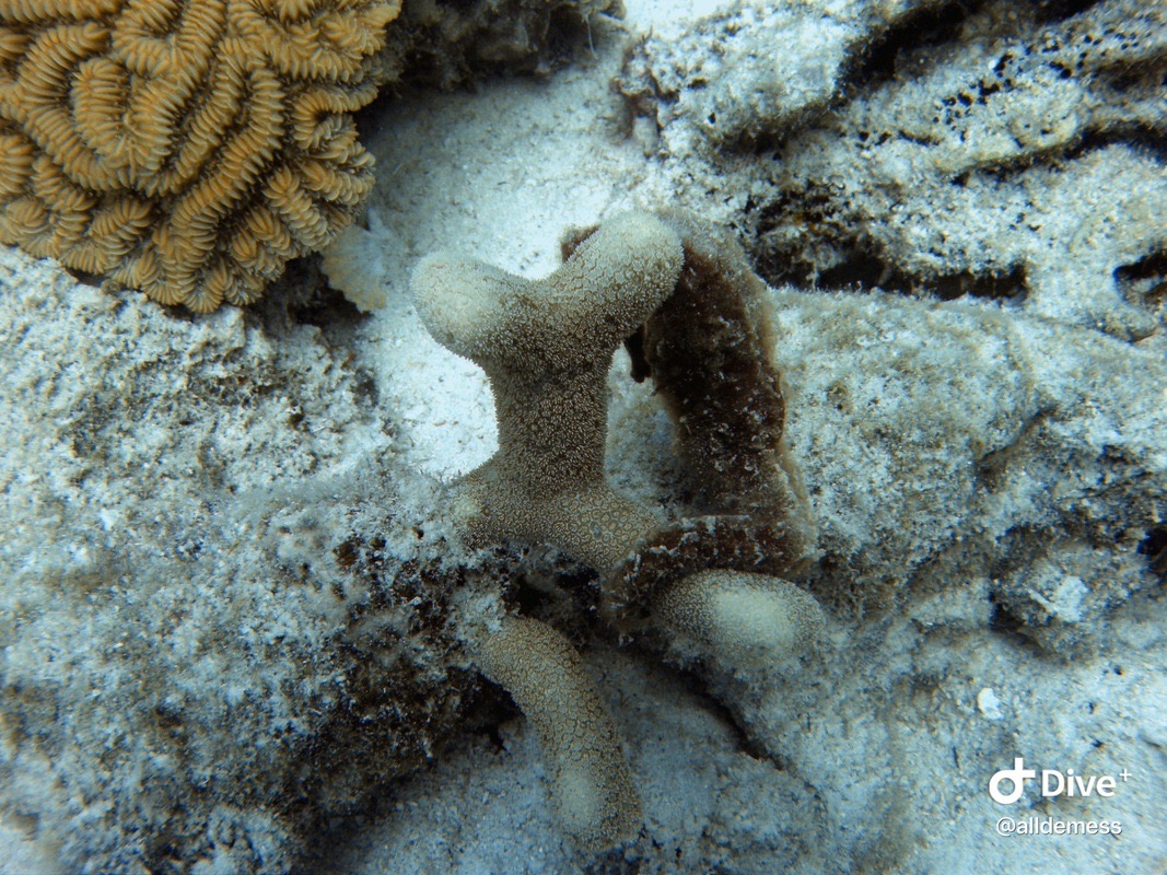
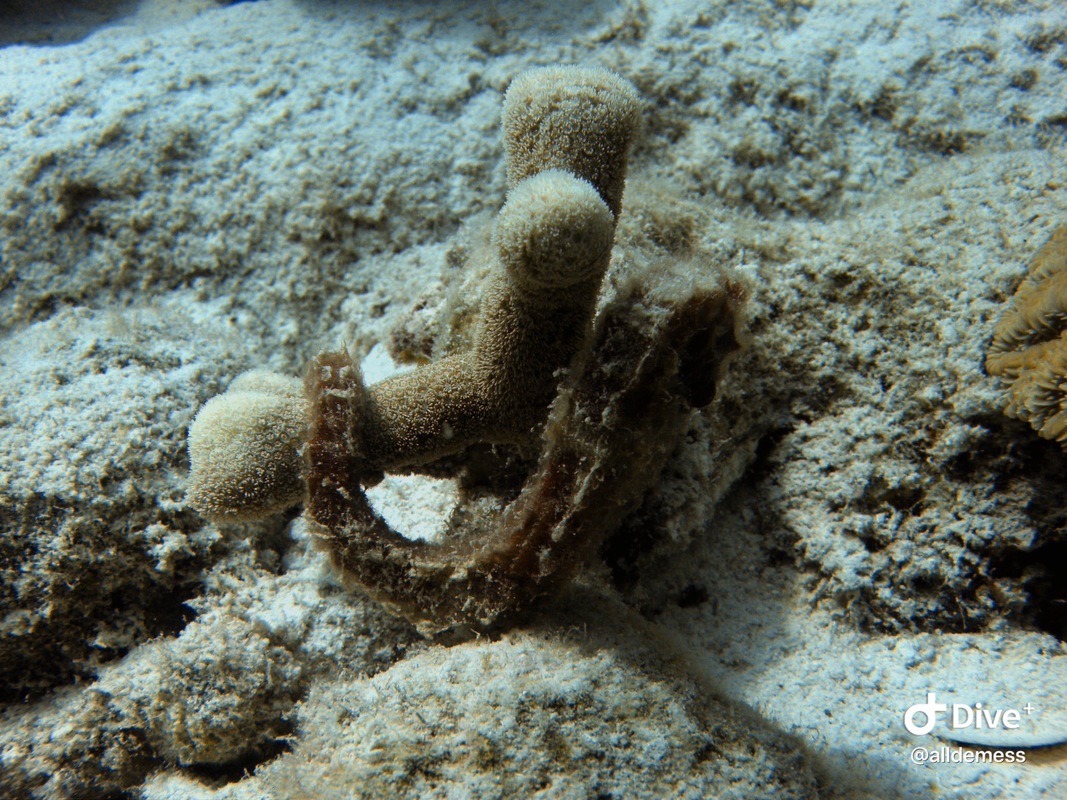
Acropora cervicornis (staghorn coral) This picture depicts two juvenile corals that settled next to each other. This ridge is the two distinct genetic entities competing for space with each other, and recognizing their non-selves!
Acropora cervicornis (staghorn coral)
Acropora cervicornis (staghorn coral)
Acropora cervicornis (staghorn coral)
Acropora cervicornis (staghorn coral)
Acropora cervicornis (staghorn coral)
Acropora cervicornis (staghorn coral)
Acropora cervicornis (staghorn coral)
Acropora cervicornis (staghorn coral)
Acropora cervicornis (staghorn coral)
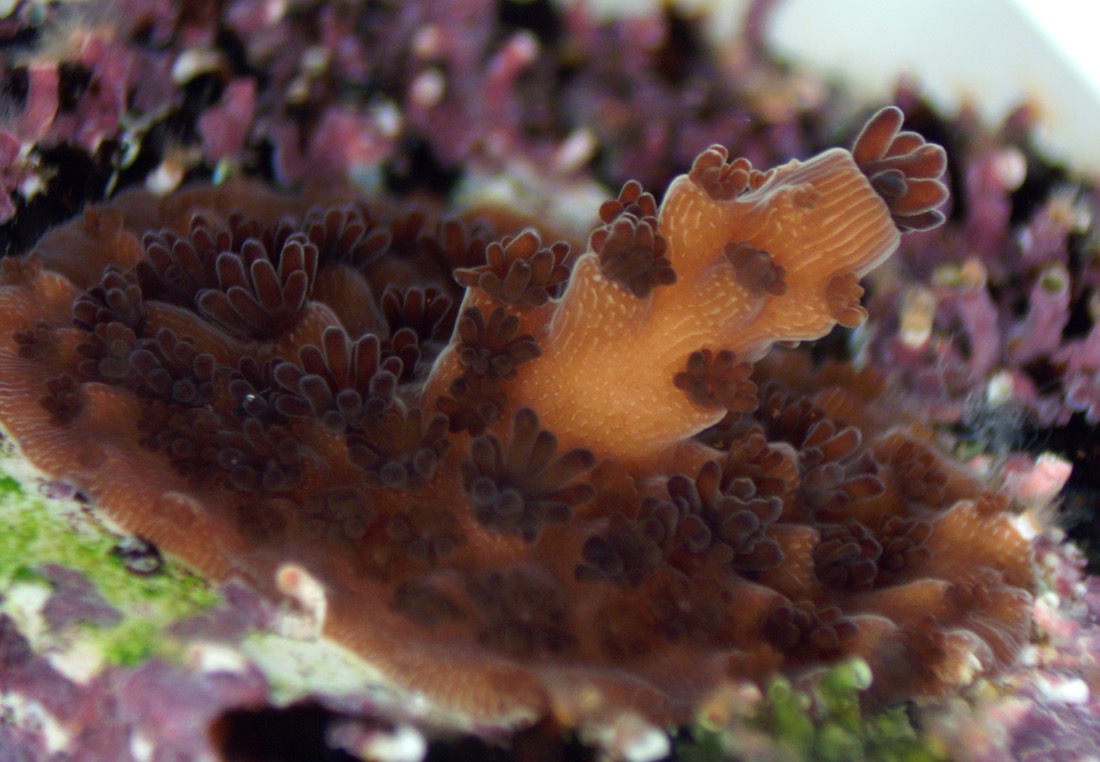
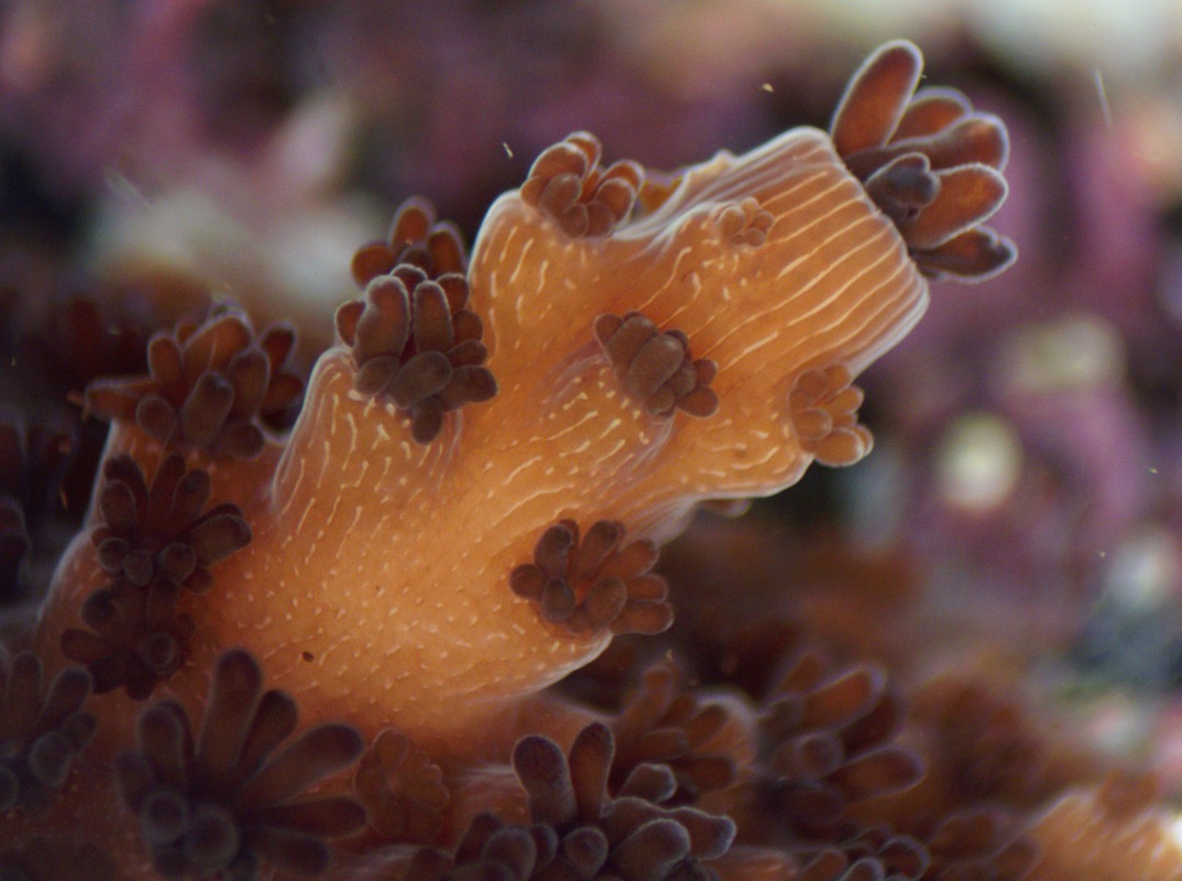
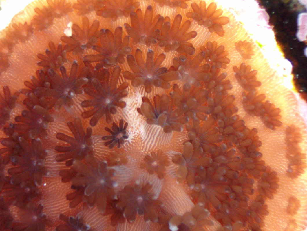
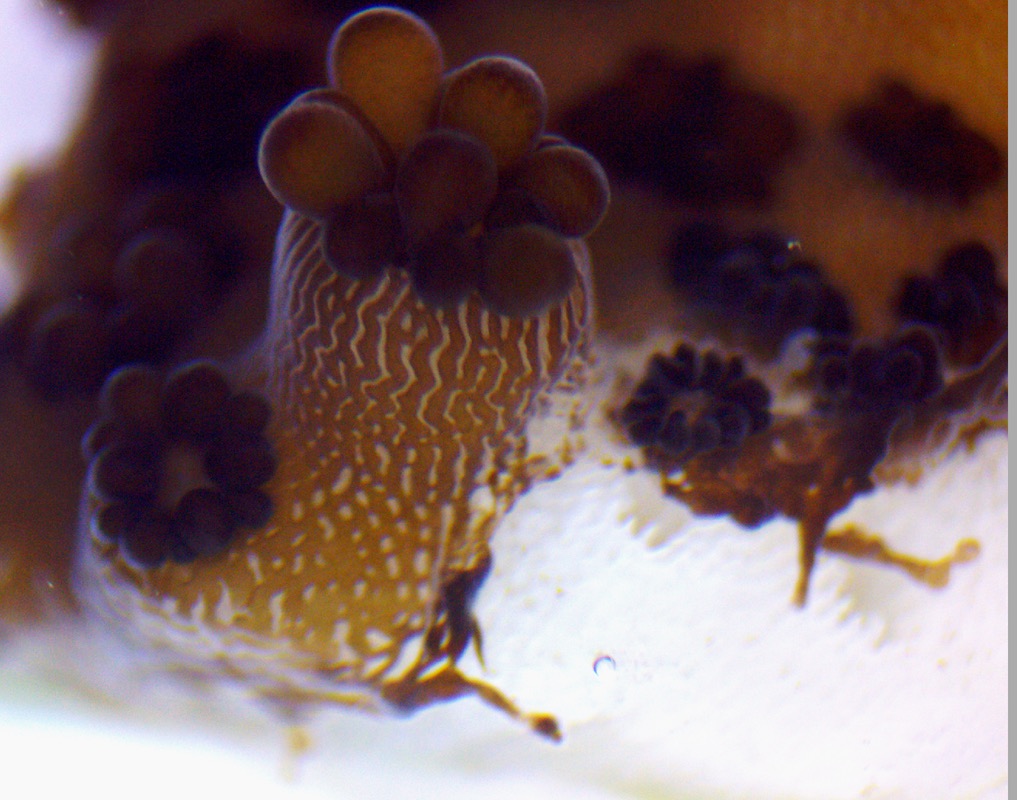
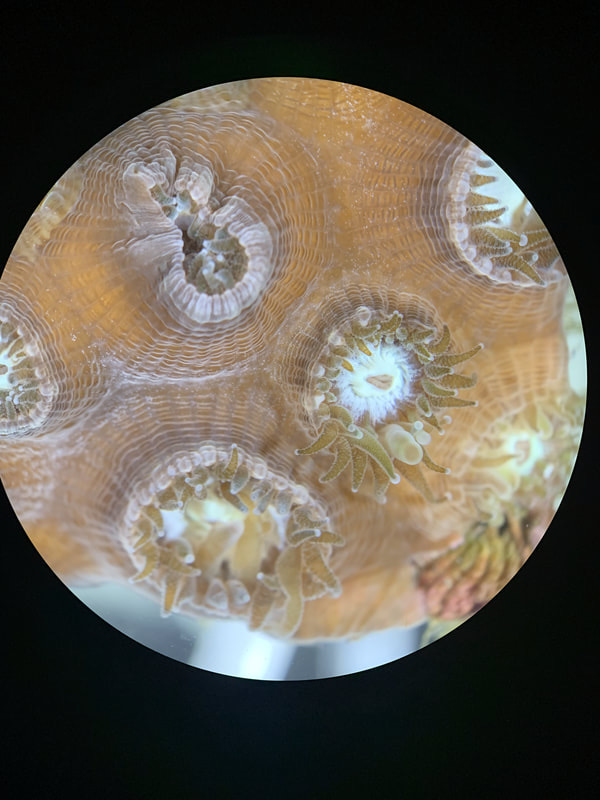
Acropora cervicornis (staghorn coral). You can see signs of stress in the center where there are white striations in the coral tissue. The coral’s mesenterial filaments (stomach lining) are protruding, which is usually a sign of stress.
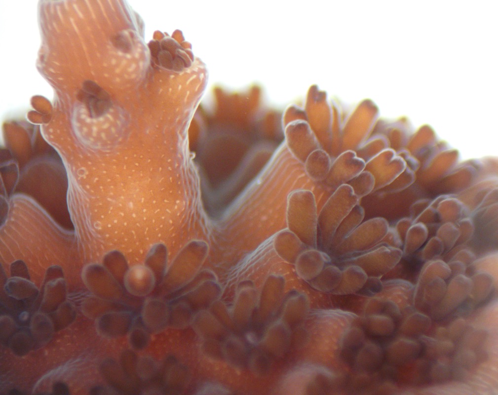
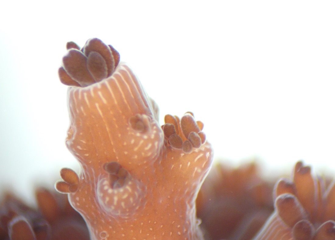
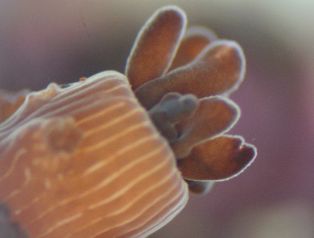
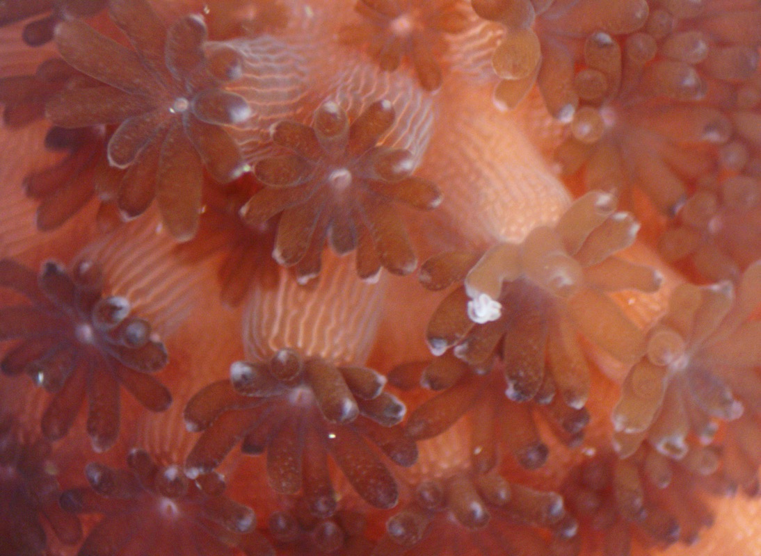
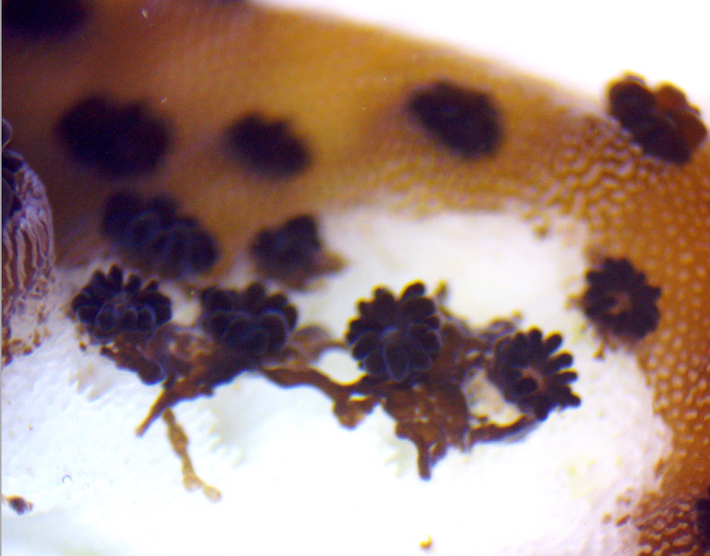
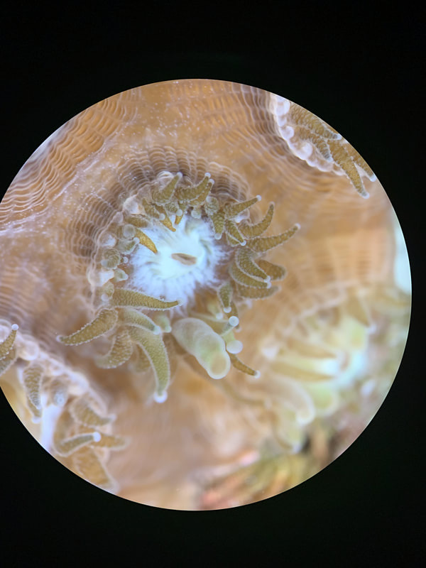
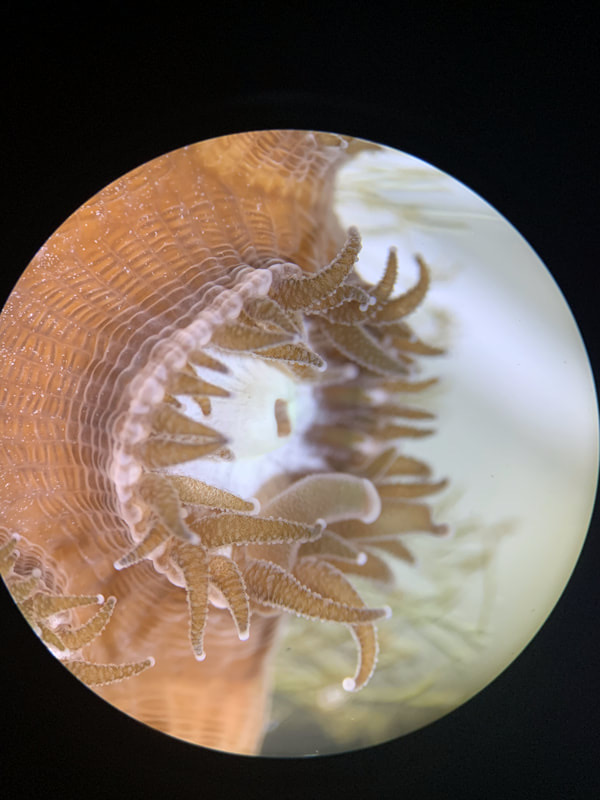
Acropora cervicornis (staghorn coral). A close-up of the mesenterial filaments emerging from the tips of the coral’s tentacles. This filaments are also used for feeding and defense.
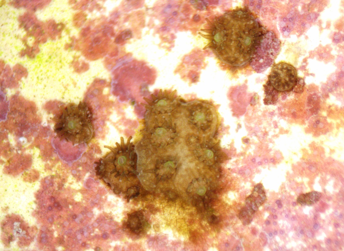
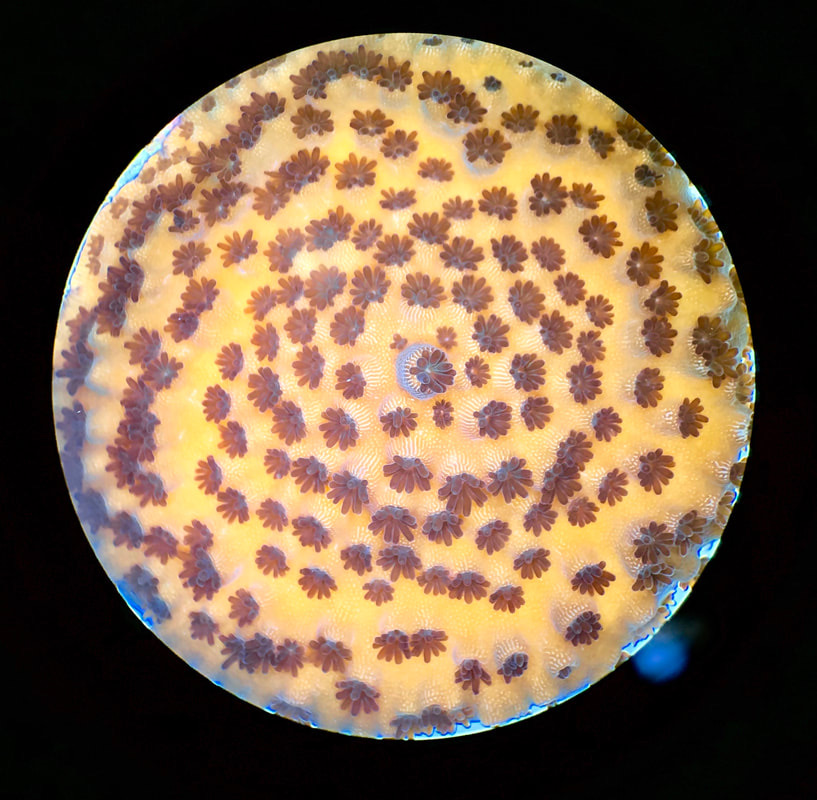
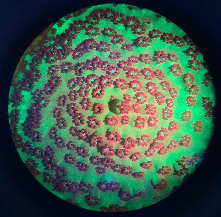
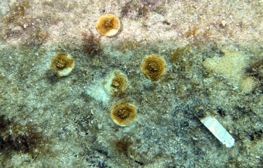
Porites astreoides (Mustard hill coral). There are several juvenile corals that have settled here on crustose coraline algae (CCA, in purple), which is a positive settlement cue for the corals. At the bottom of the picture you can see a larger coral growing over top of a smaller coral. Another recognition of non-self and competition for space!
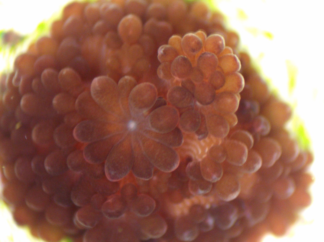
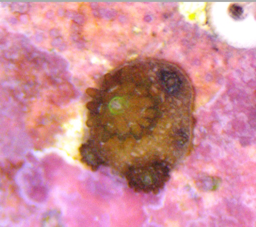
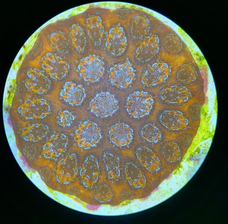
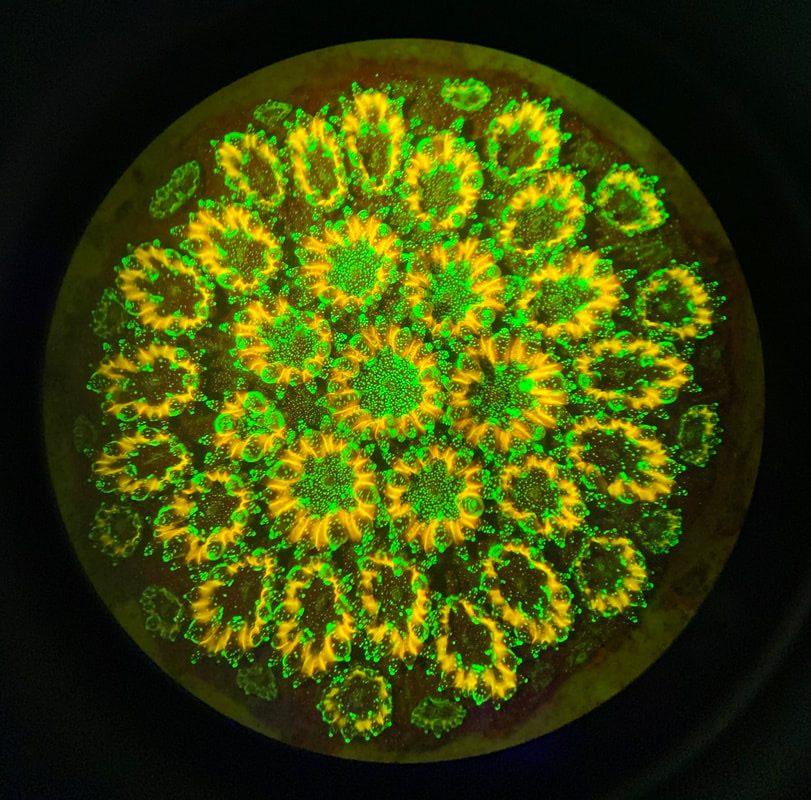
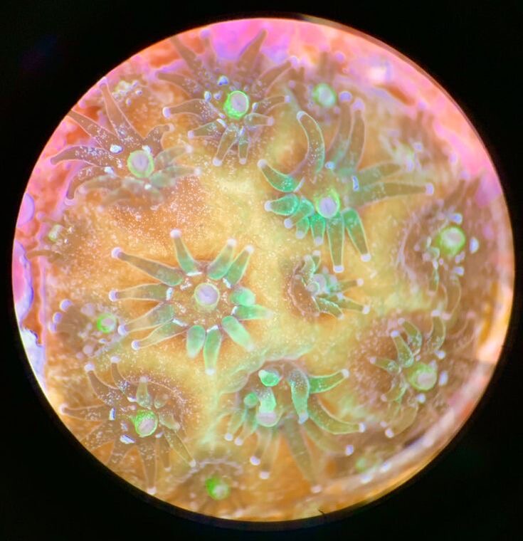
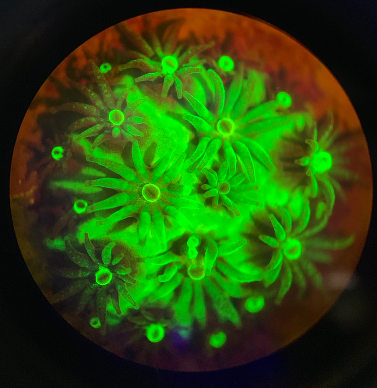
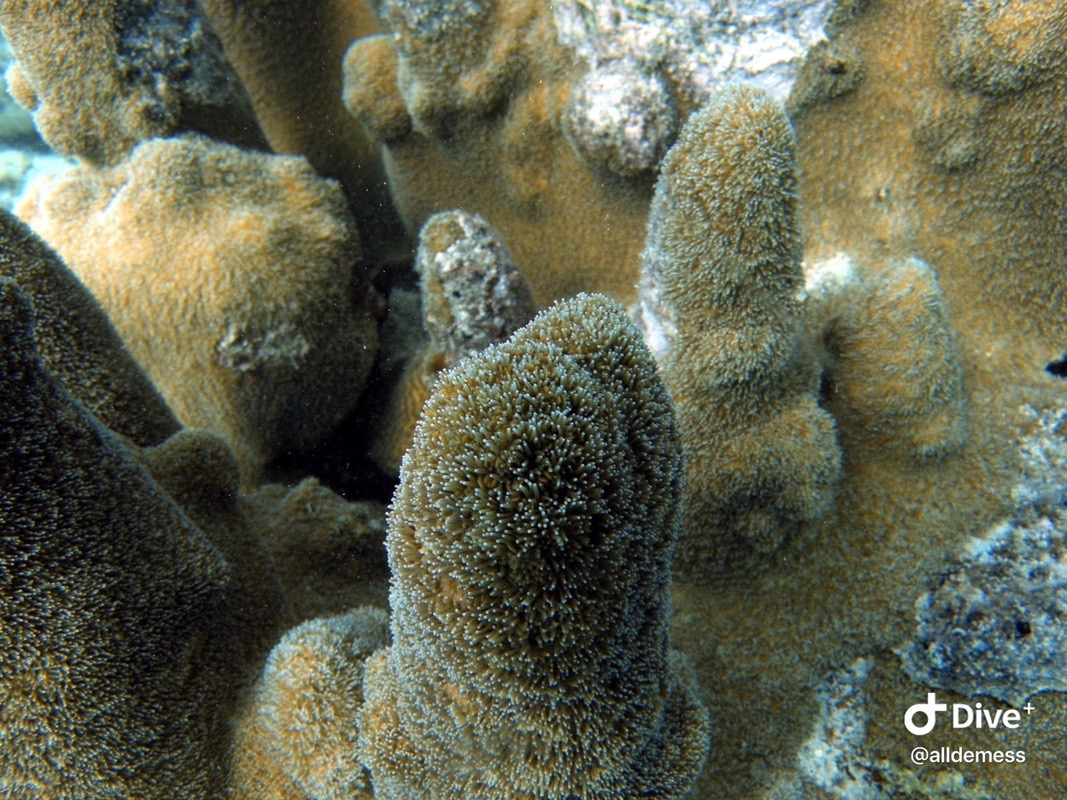
Porites astreoides (Mustard hill coral). One coral juvenile with its central polyp (mouth in green), and growing tiny polyps circling it.
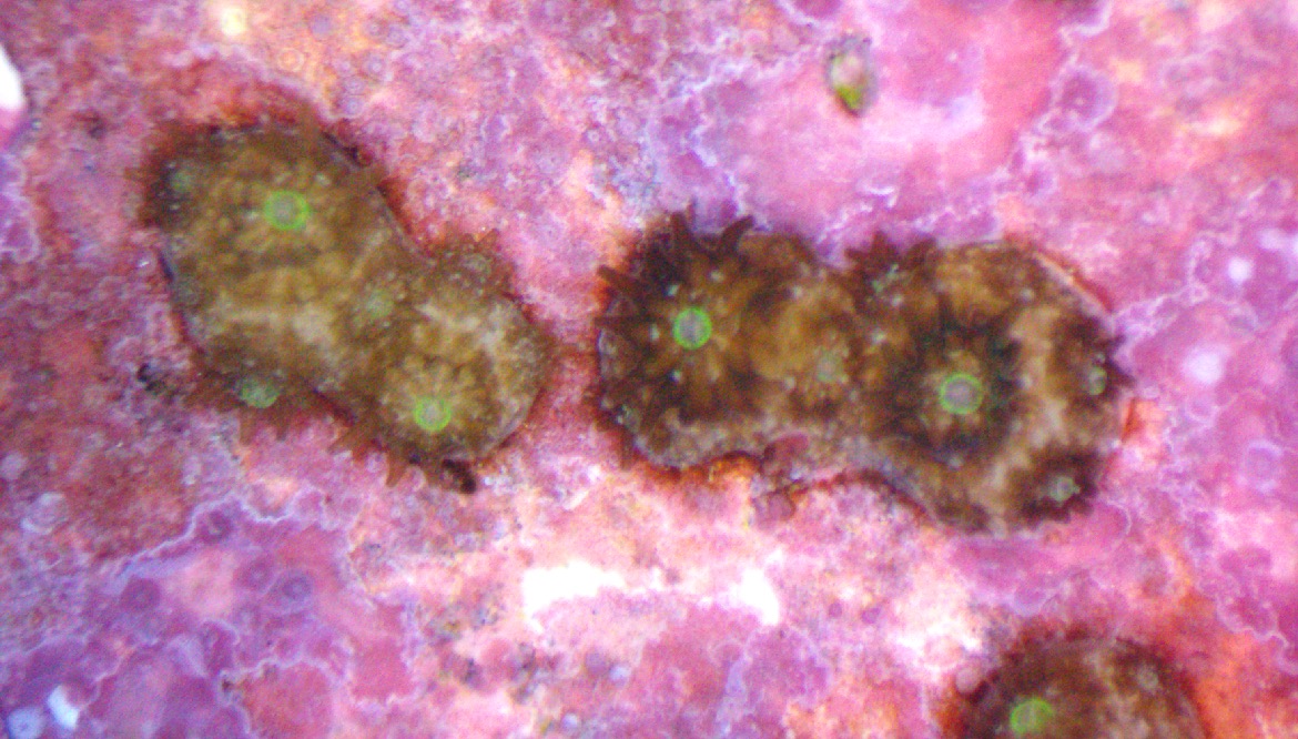
Porites astreoides (Mustard hill coral). These juvenile corals are also distinct genetic entities, but are actually fusing together instead of competing for space! Researchers are still trying to understand why this occurs for some corals and not others. Google
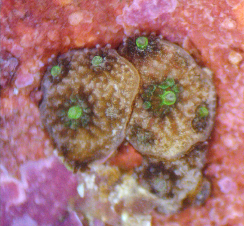
Porites astreoides (Mustard hill coral). Two juvenile corals competing for space as they grow.
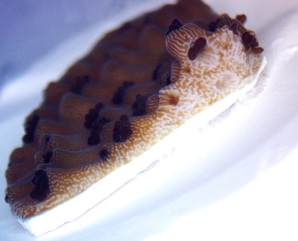
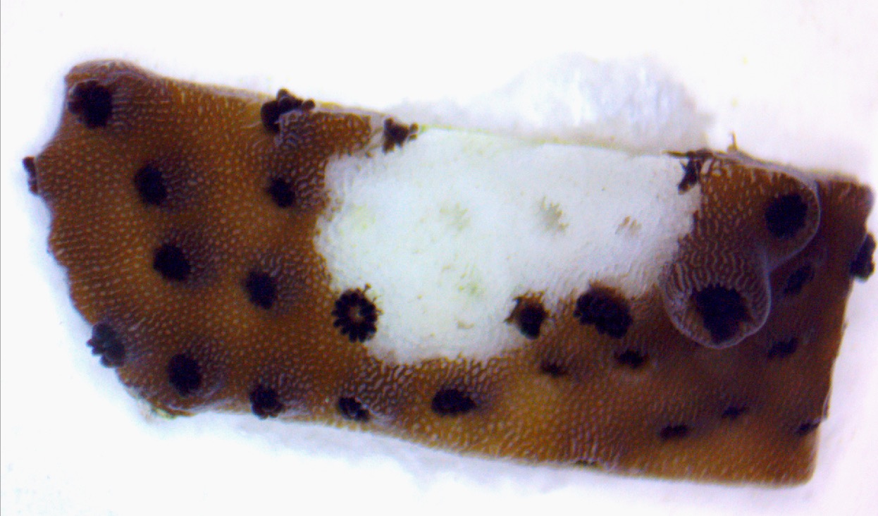
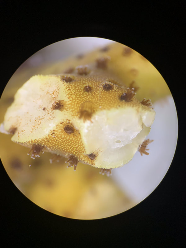
Acropora cervicornis (staghorn coral). This is a juvenile coral right after being micro-fragmented. Micro-fragmentation is a restoration technique designed by Mote Marine Laboratory’s Dr. David Vaughan.
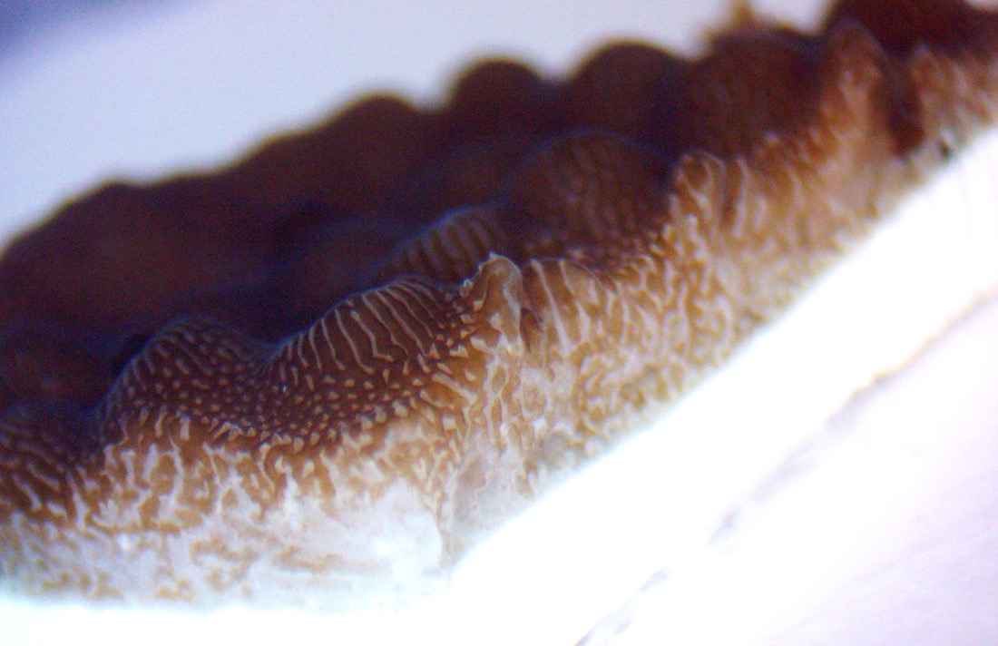
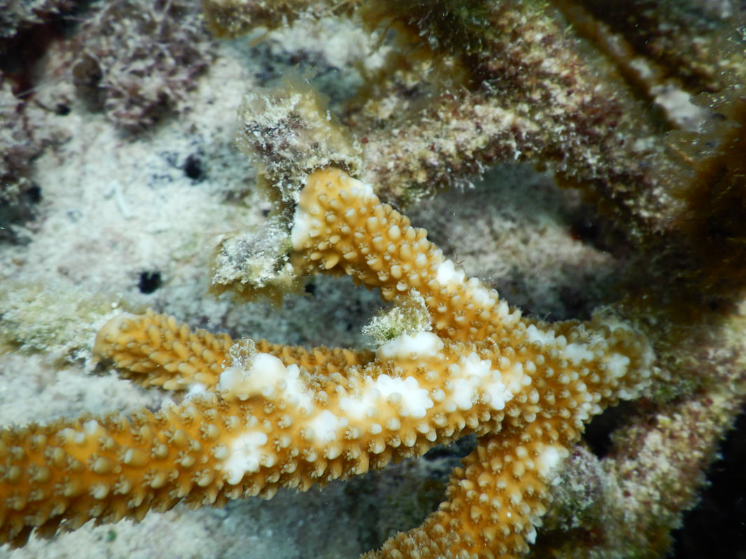
Acropora cervicornis (staghorn coral). You can see the freshly cut coral tissue along the white line.
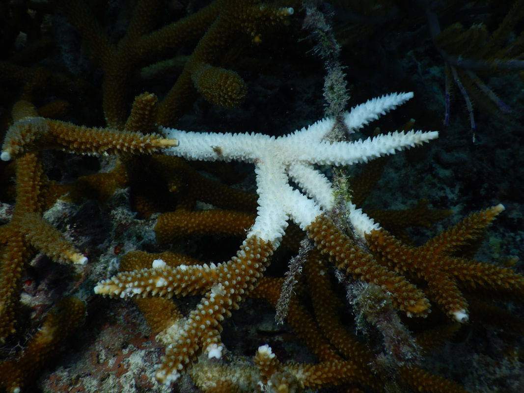
Acropora cervicornis (staghorn coral). This coral fragment, days after being micro-fragmented, got really stressed and started sloughing, or releasing, its tissue. We are not sure why this happened to this fragment and not others - it was clearly stressed out.
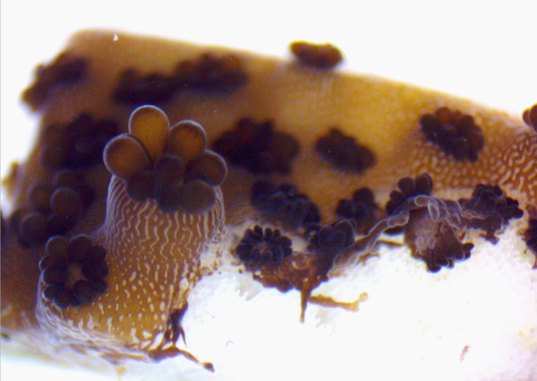
Acropora cervicornis (staghorn coral). Another close-up of the tissue sloughing; it is interesting to me to see the connectedness of the tentacles and tissue layer, with the white calcified skeleton underneath.
Acropora cervicornis (staghorn coral)
Acropora cervicornis (staghorn coral). A top-down view of that same coral fragment after the tissue was fully released. You can see the white coral skeleton and the indents for where the polyps sat.
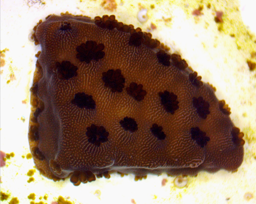
Acropora cervicornis (staghorn coral). This coral fragment, on the other hand, fared much better and is growing past its cuts! You can see the thin layer of tissue extending and growing new polyps with tentacles as it continues to grow.
Acropora cervicornis (staghorn coral). More coral growth!
Acropora cervicornis (staghorn coral). You can see the faint change from dark brown to light brown on the side; this is where the coral tissue has grown and extended down the side of the fragment, and has now grown new polyps!
Acropora cervicornis (staghorn coral)
Acropora cervicornis (staghorn coral). After growing outwards, its time for the coral to start growing upwards! It is a branching coral after all.
Acropora cervicornis (staghorn coral)
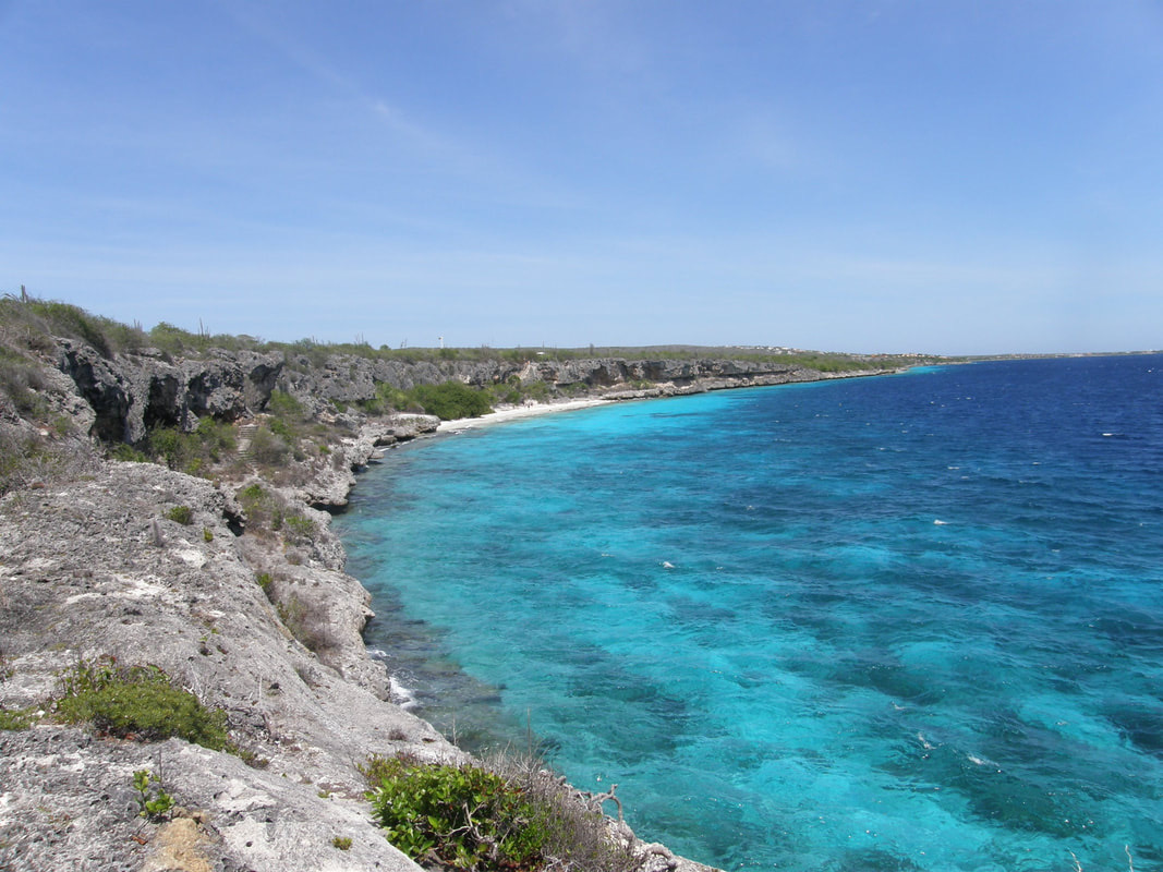
Beautiful coastline of Bonaire
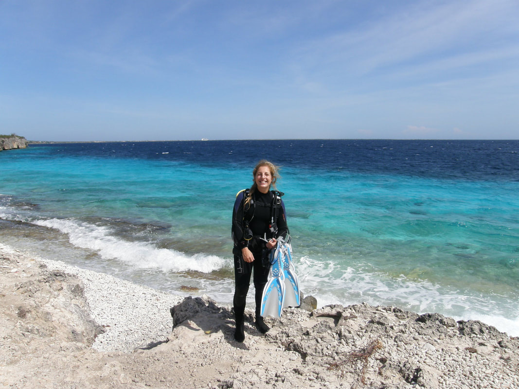
I’m ready to go shore-diving!
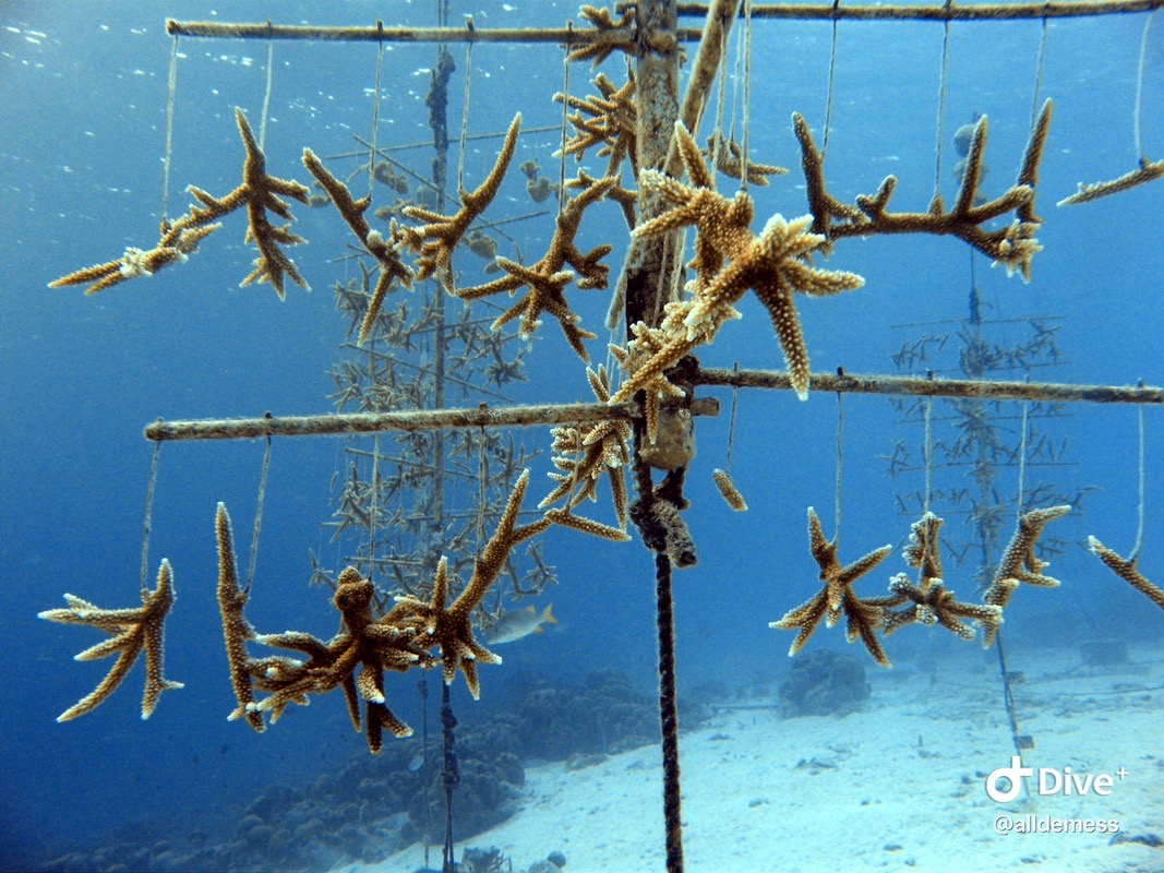
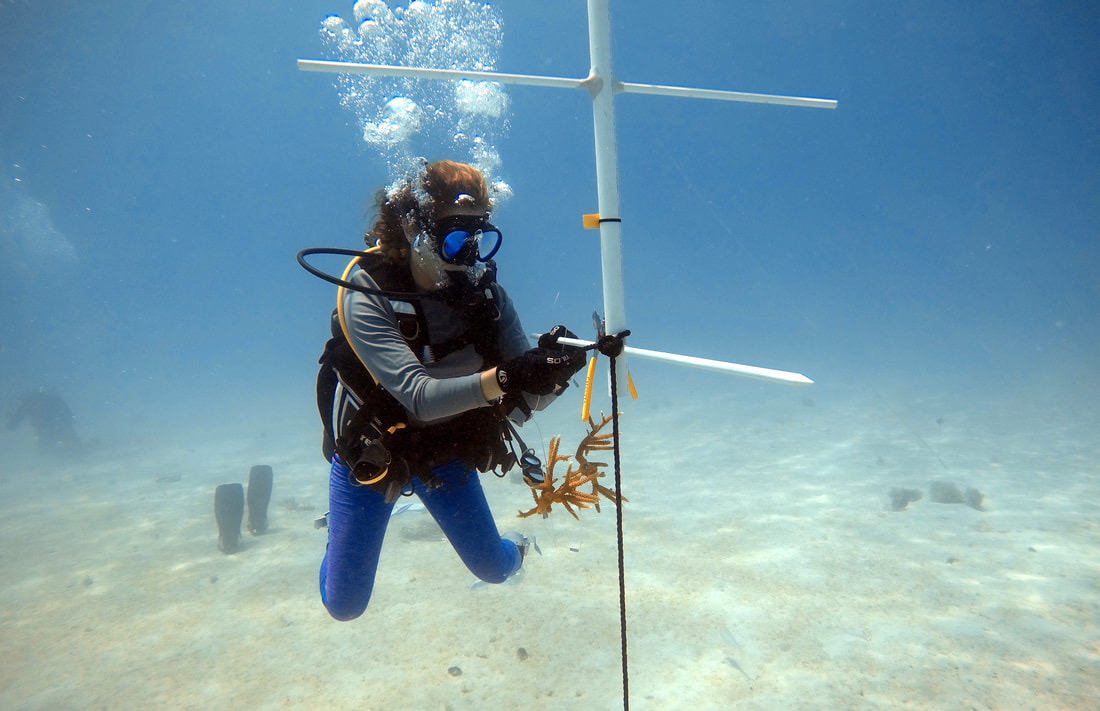
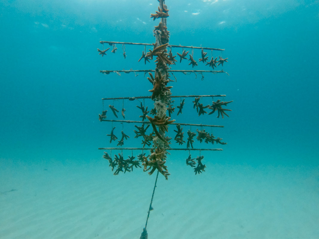
One of Reef Renewal’s nursery trees for Acropora cervicornis (staghorn coral)
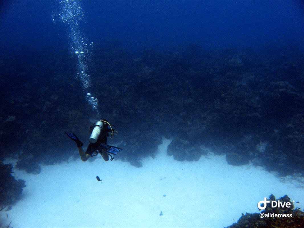
Kristin going over the reef wall!
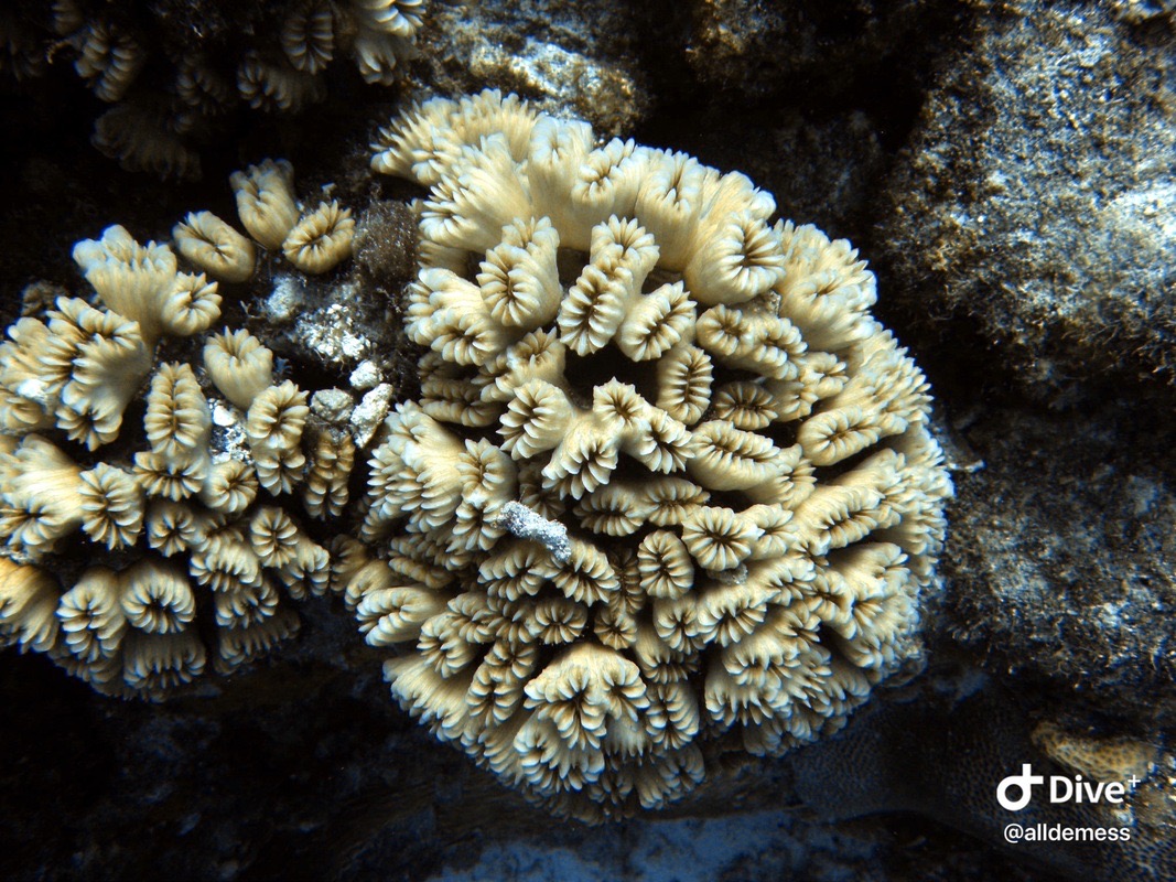
Eusmilia fastigiata (smooth flower coral)
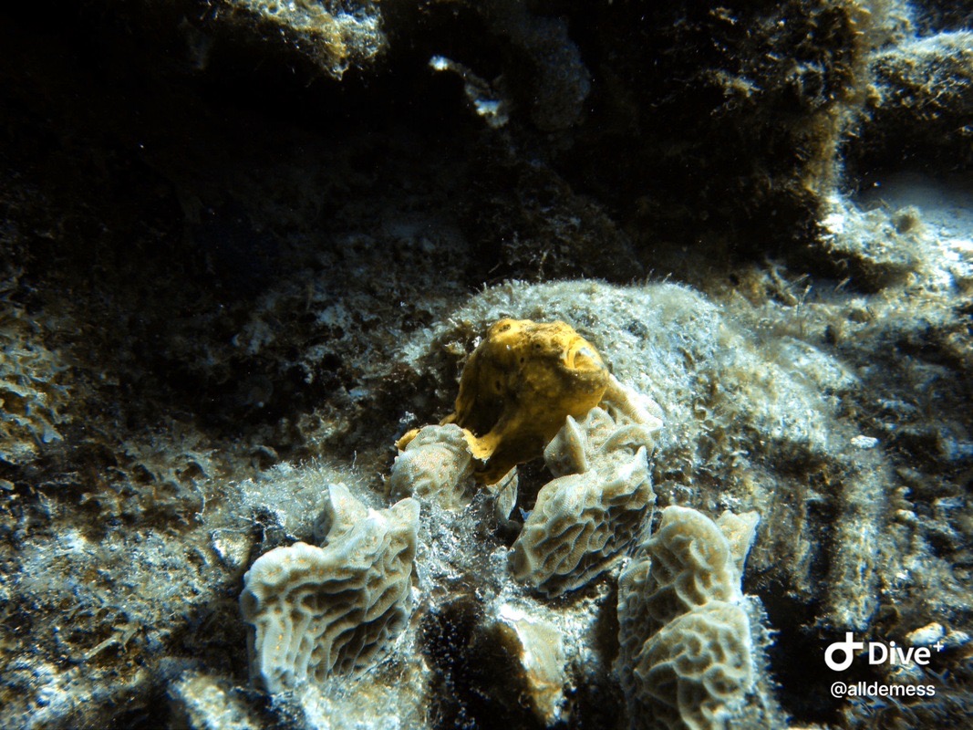
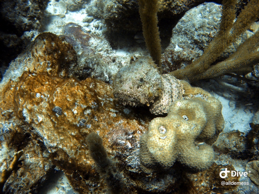
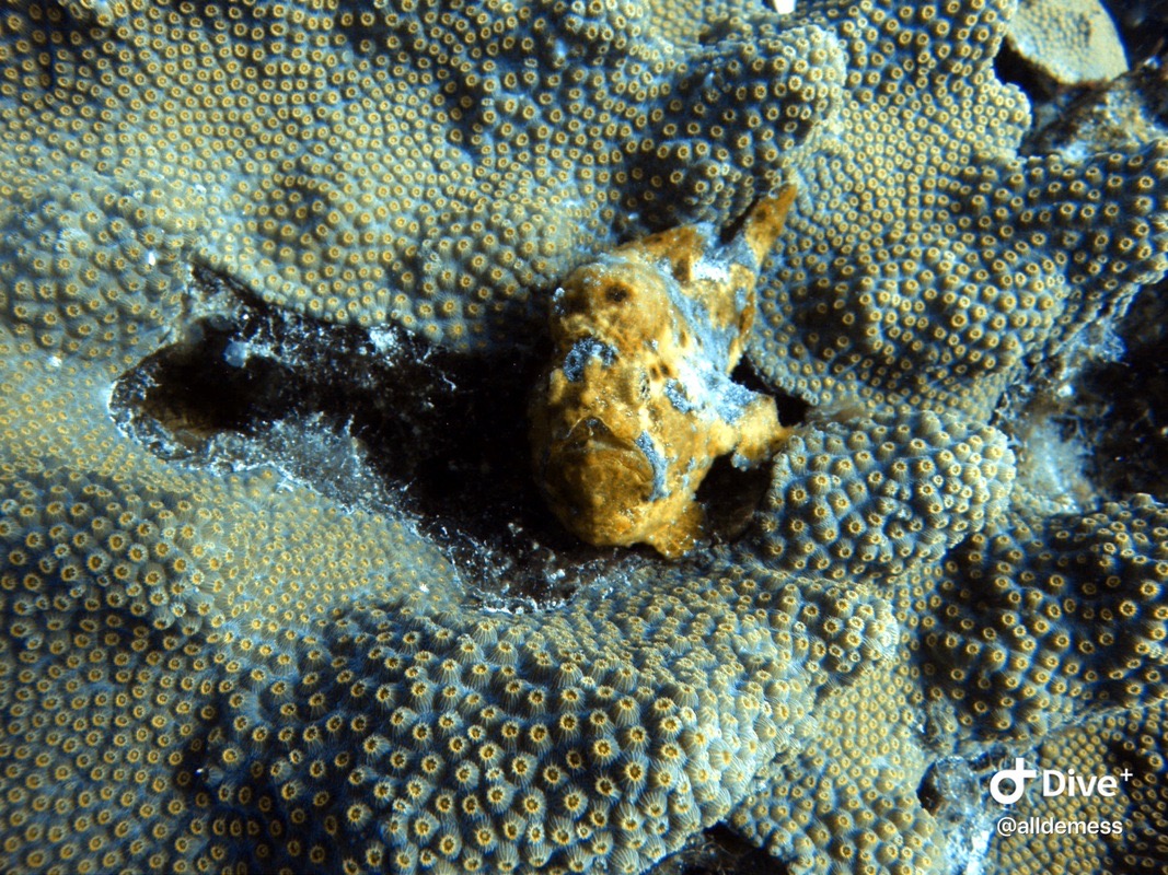
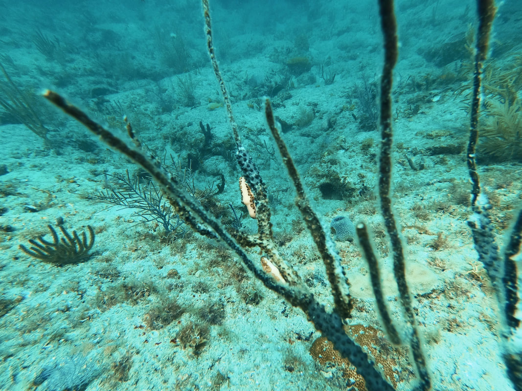
A frogfish! These are so fun (and challenging) to find because they can blend in super well with sponges and corals. This one was thankfully failing at camouflaging.
Josh and Kristin at Captain Don’s memorial site
Kristin and the reef wall (depth ~70 ft)
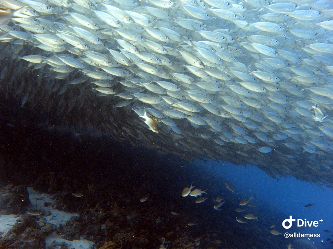
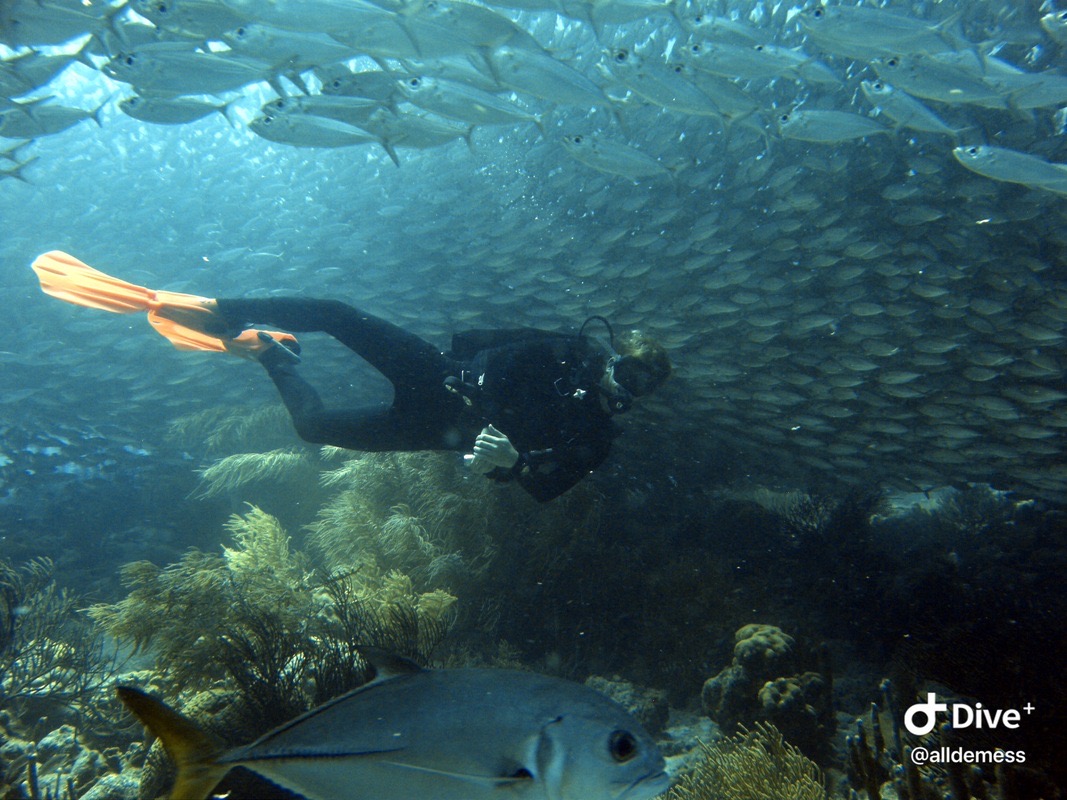
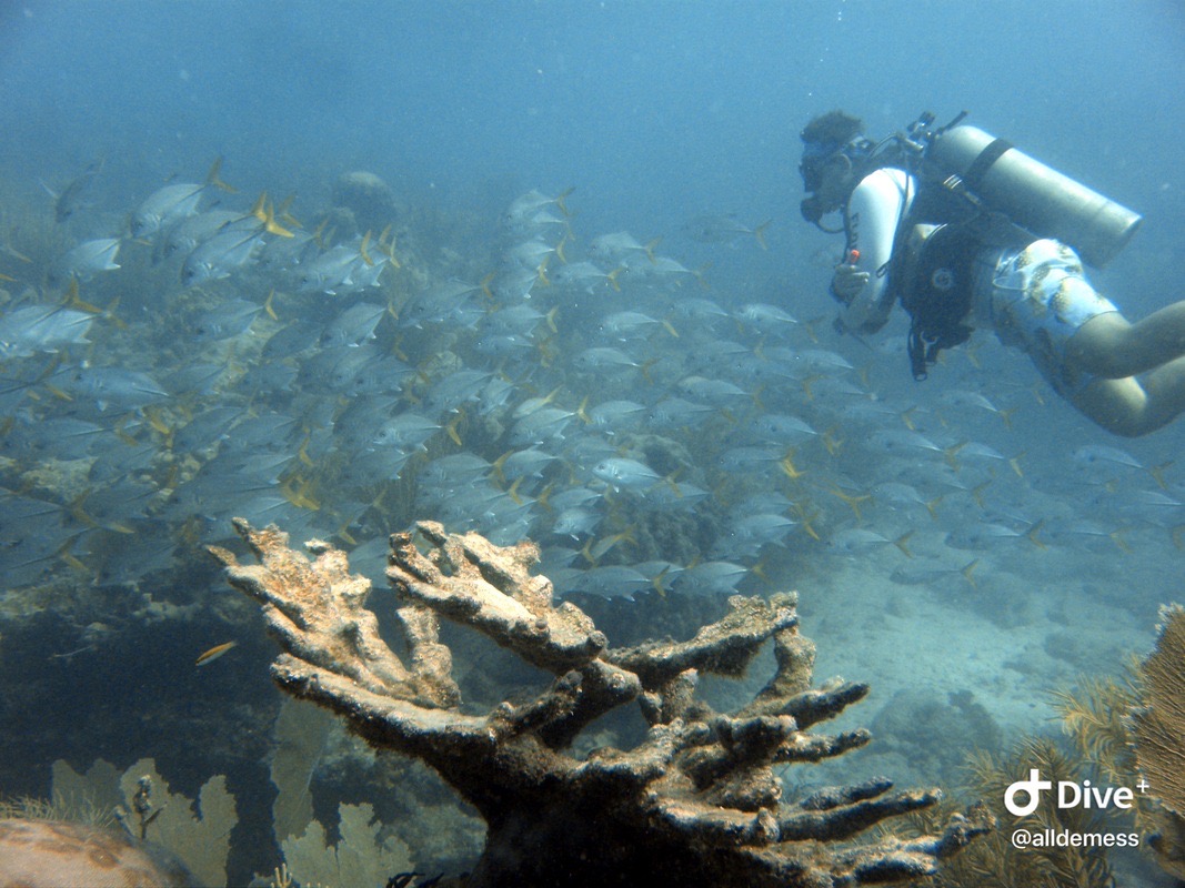
A bait ball full of fish!
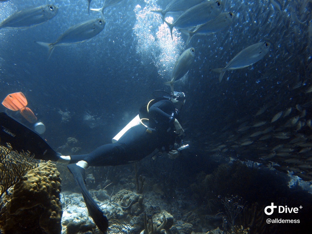
Naturally we swam through it. Kristin in the middle of the bait ball!
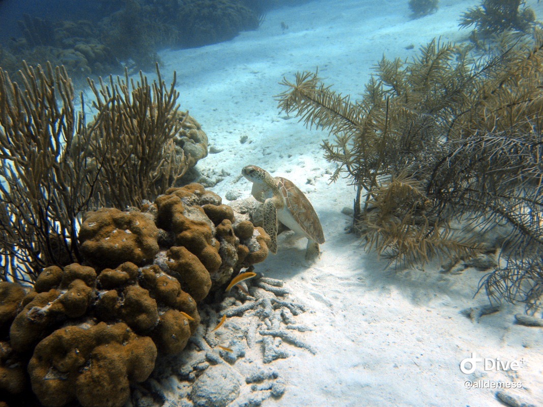
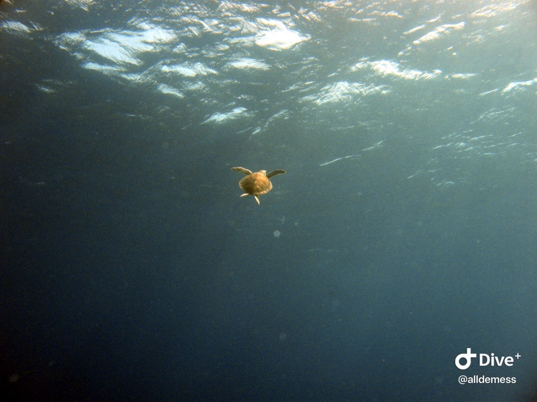
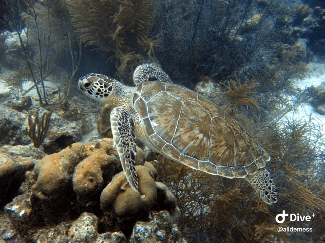
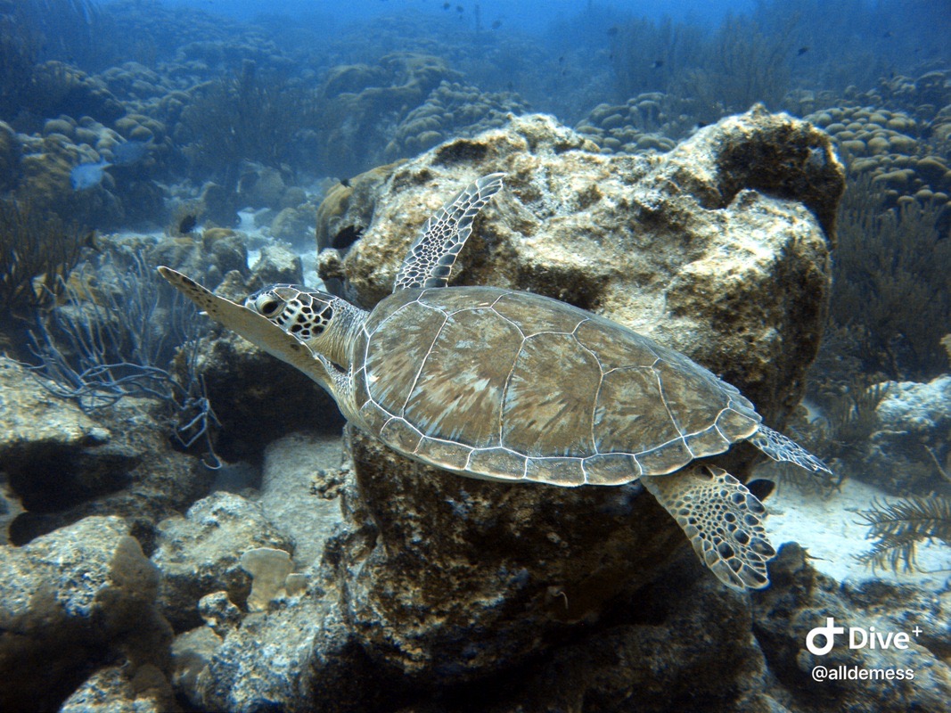
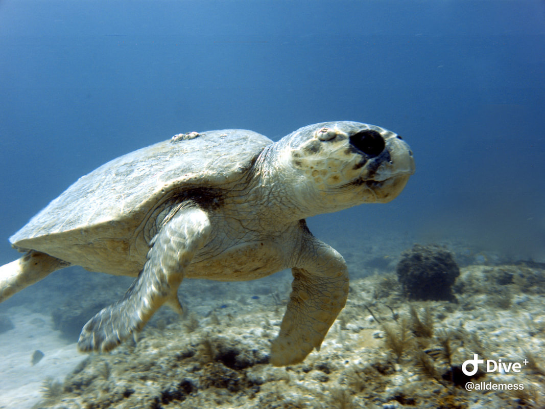
A juvenile sea turtle!!! My favorite sea creature to see while diving.
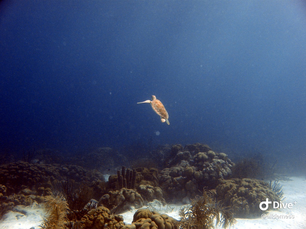
He did not want to hang with us.
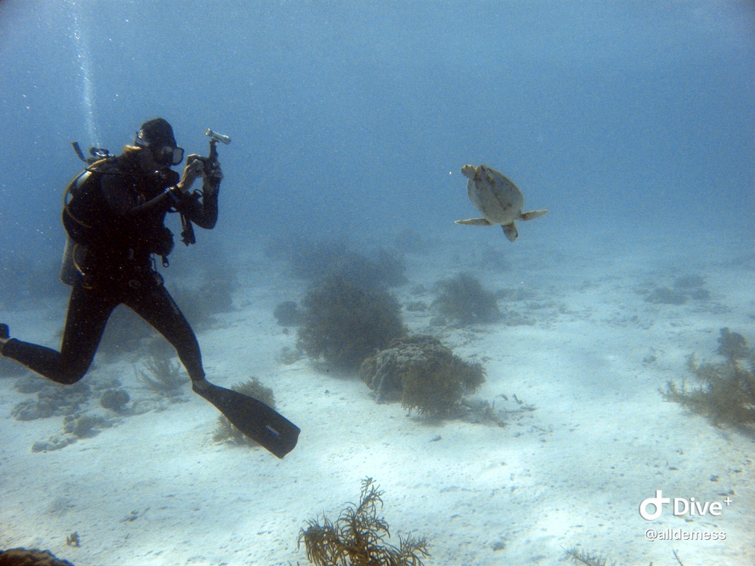
He did swim by Kristin so she could get a cool pic.
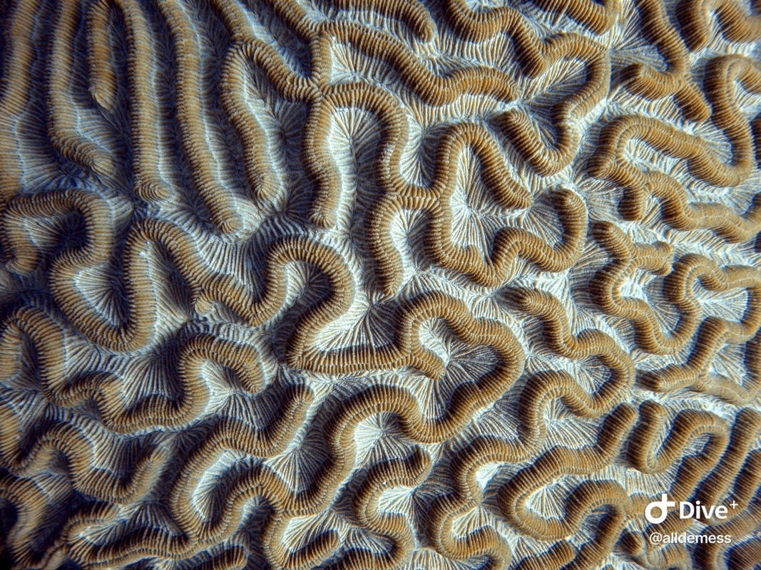
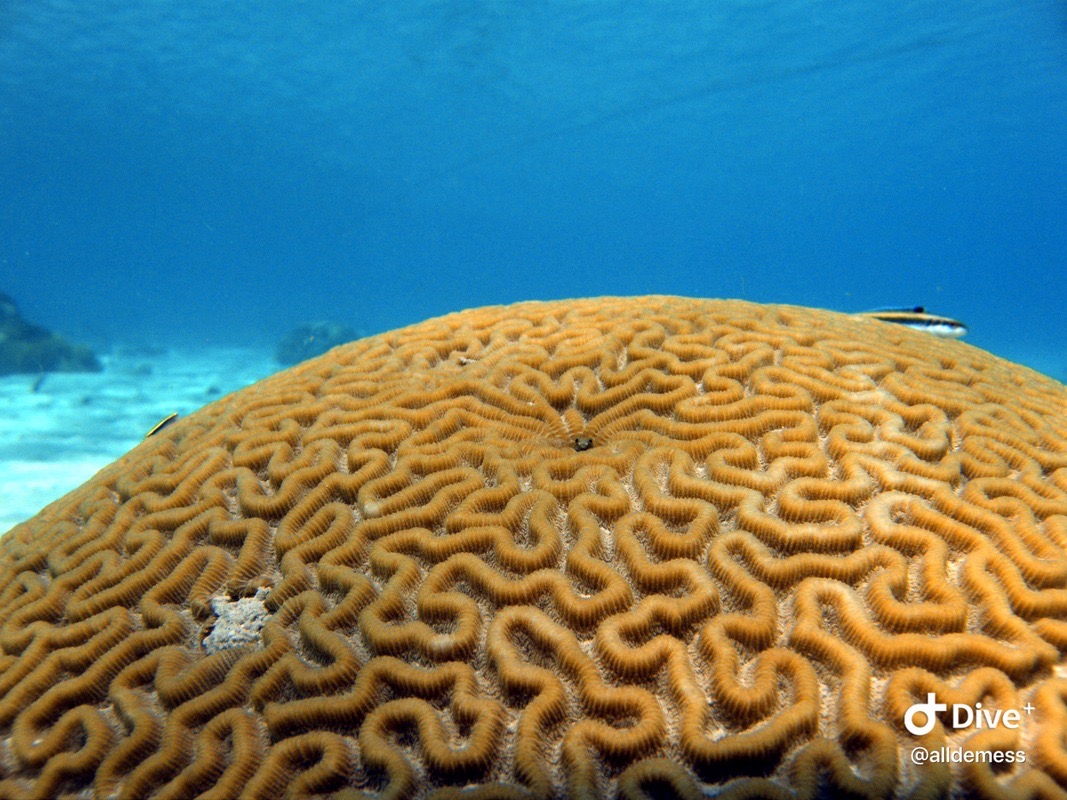
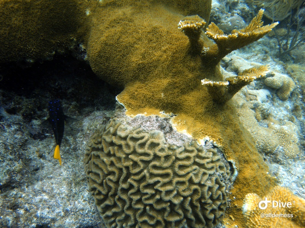
Pseudodiploria strigosa (brain coral)
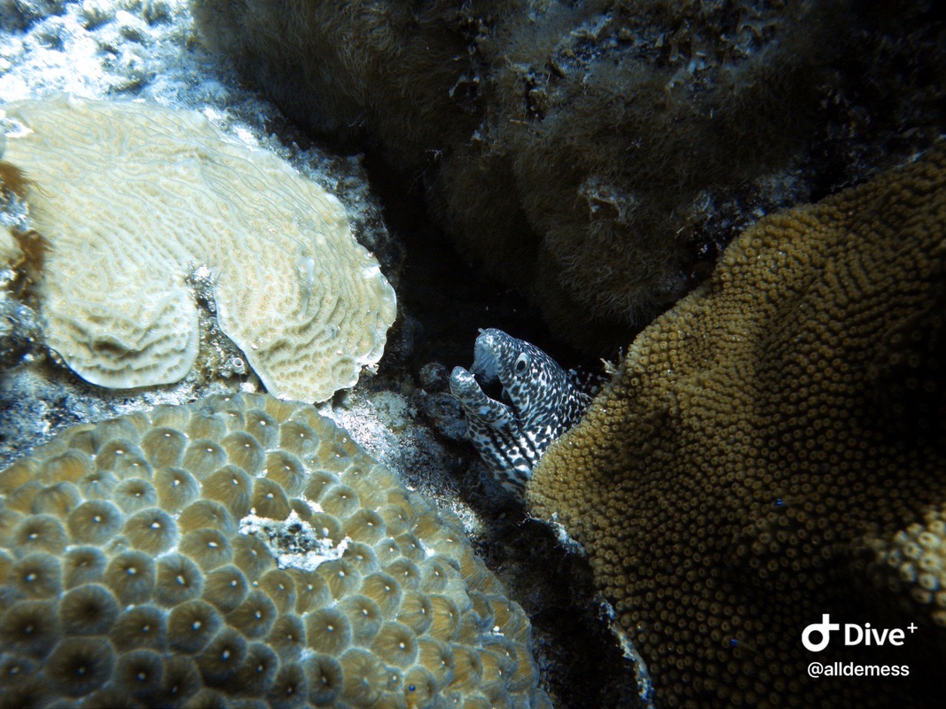
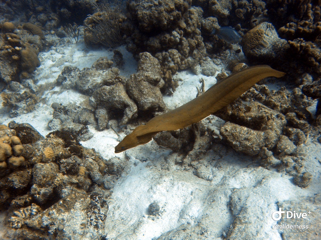
A green moray swimming by! Spooky.
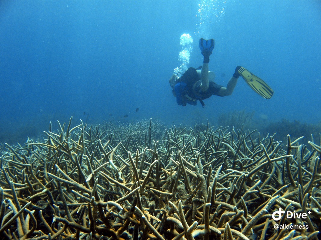
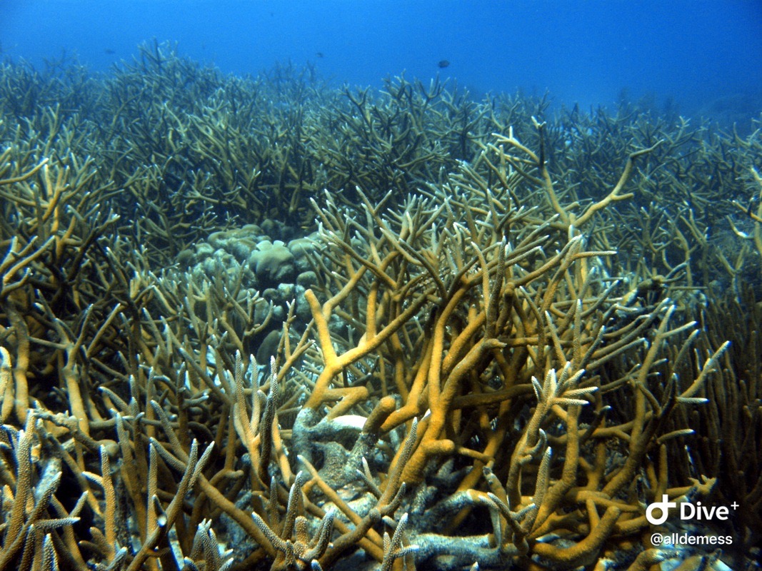
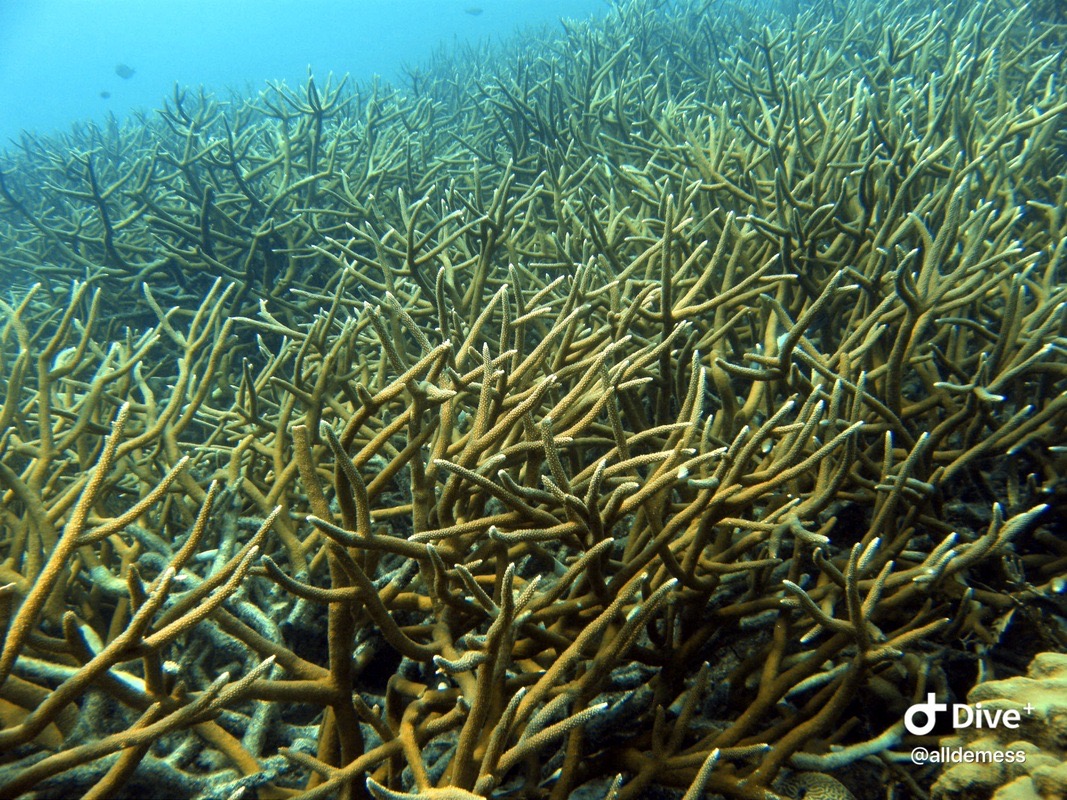
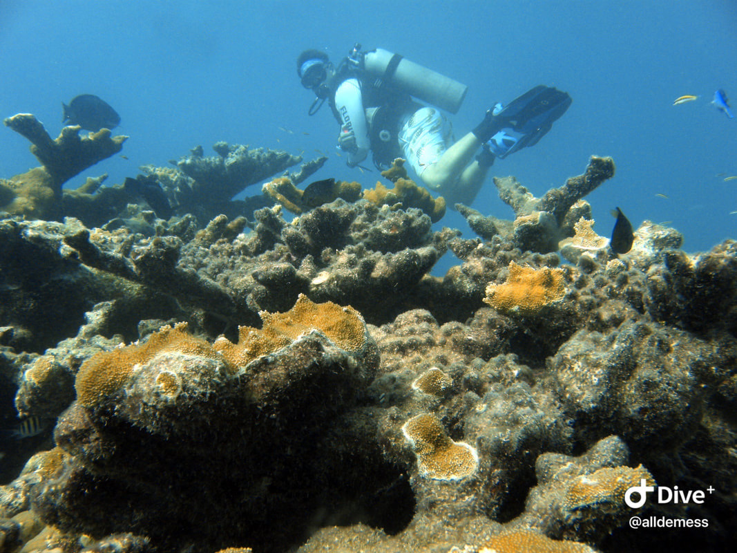
Garrett showing me the amazing natural patches of staghorn coral. I have never seen this much staghorn before!
A shy seahorse wrapped around a piece of coral.
Still so shy! I thought he was algae at first.
Another sea turtle! He hung out with us a bit more.
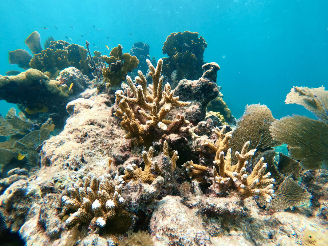
More staghorn coral patches!
Look at all that staghorn.
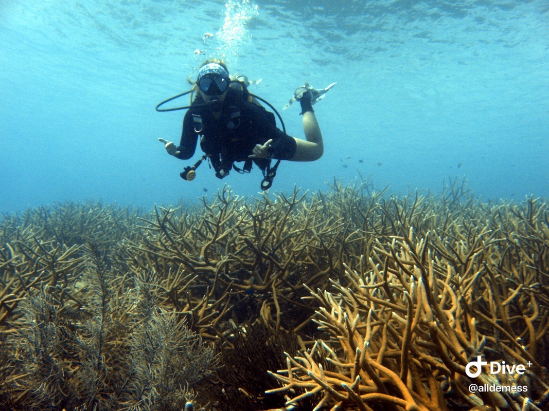
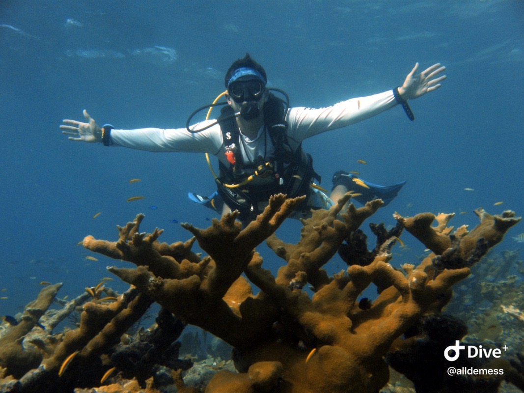
Me, super excited to be with a patch of natural staghorn coral, the species I study and am interested in helping restore in Florida.
Another frogfish that wasn’t doing the best job at hiding! He’s sitting on an Orbicella faveolata coral.
Dive site: the Lake (that sand patch in the middle, surrounded by a circular reef). Featuring Aimee Young, infamous frogfish finder!
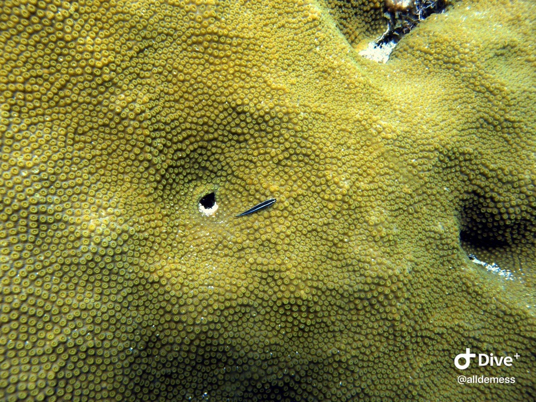
Colpophyllia natans - spot the tiny blenny fish in the middle looking at me!
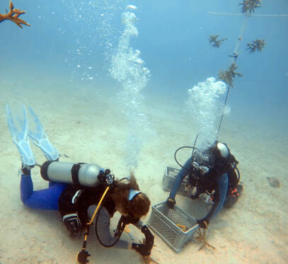
Hanging corals on their new tree! (Photo by Dr. Hanna Koch)
Myself and Sam Cook getting corals ready. (Photo by Dr. Hanna Koch)
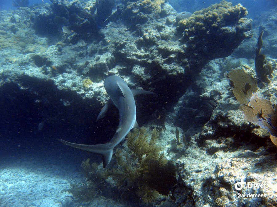
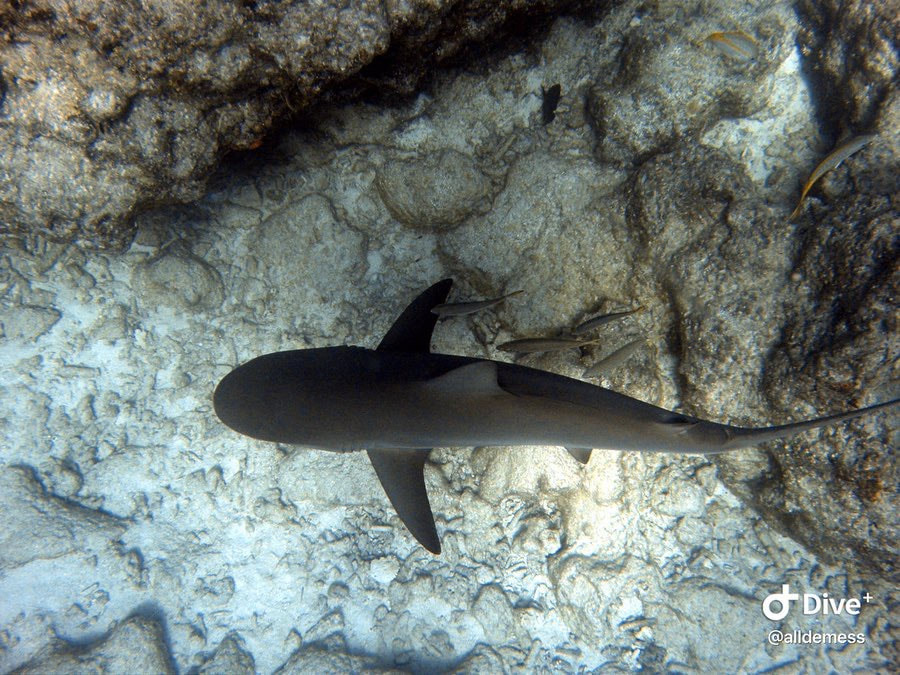
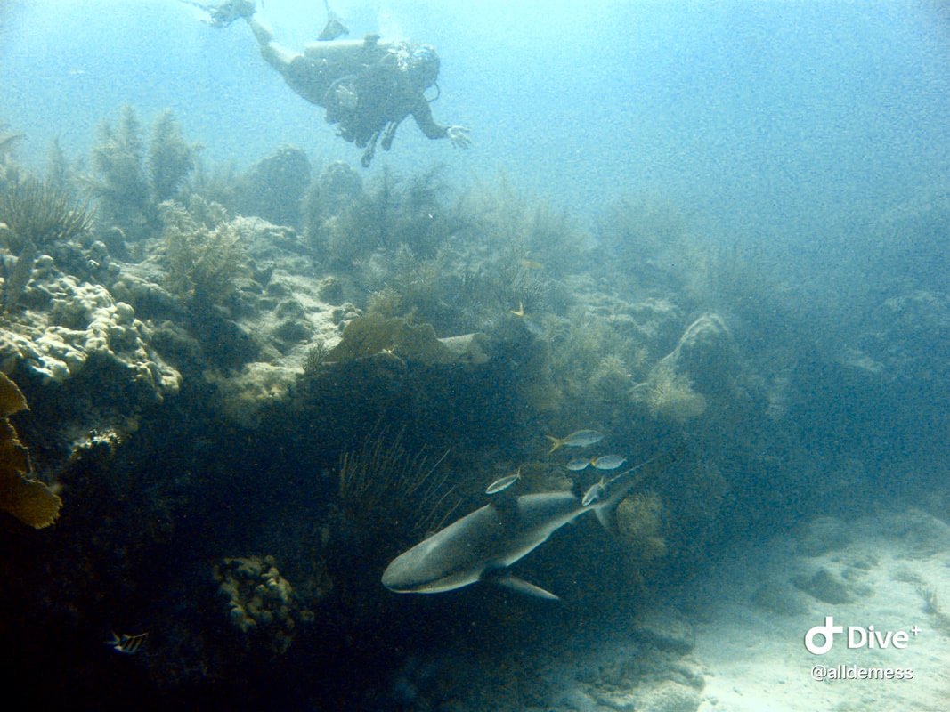
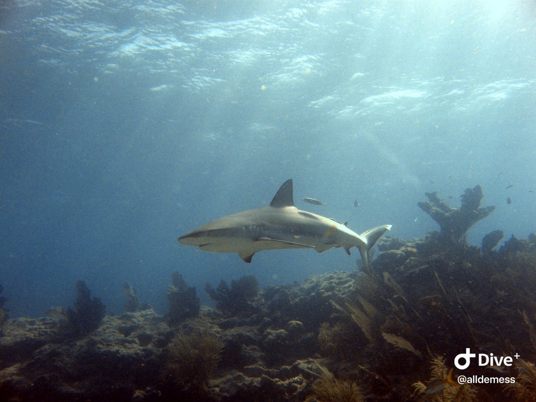
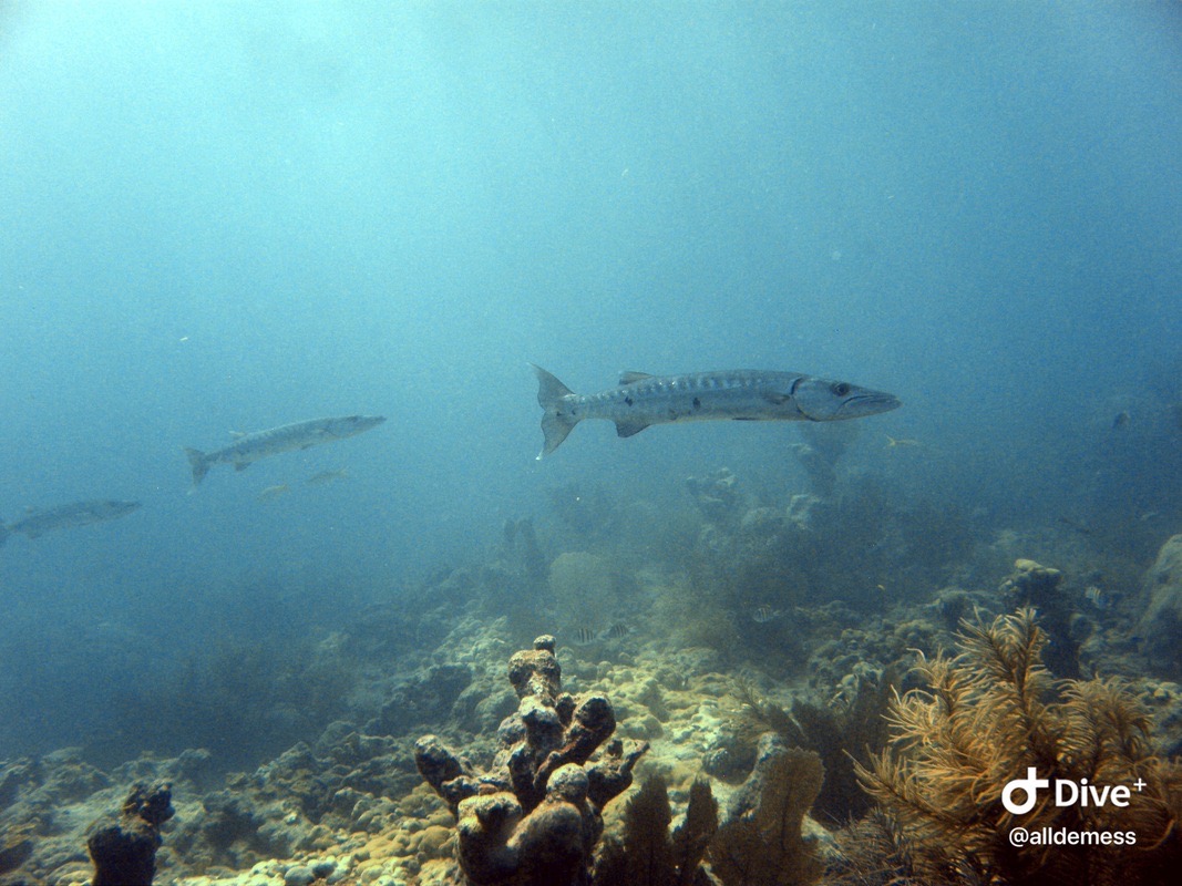
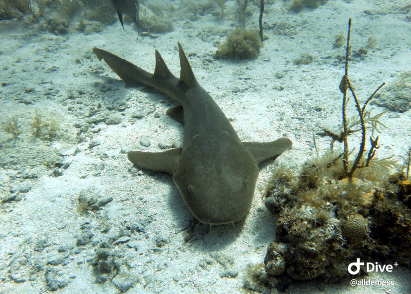
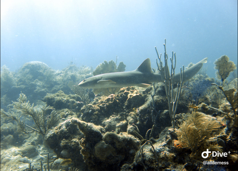
This shark was the highlight of my dive that day
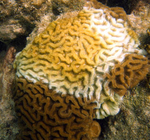
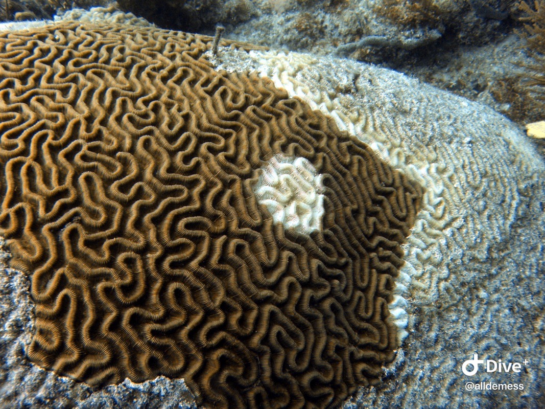
Stony coral tissue loss disease actively spreading on this brain coral. A really sad sight, but unfortunately this disease has spread throughout the Florida Keys. Learn more about the disease by clicking on the photo.
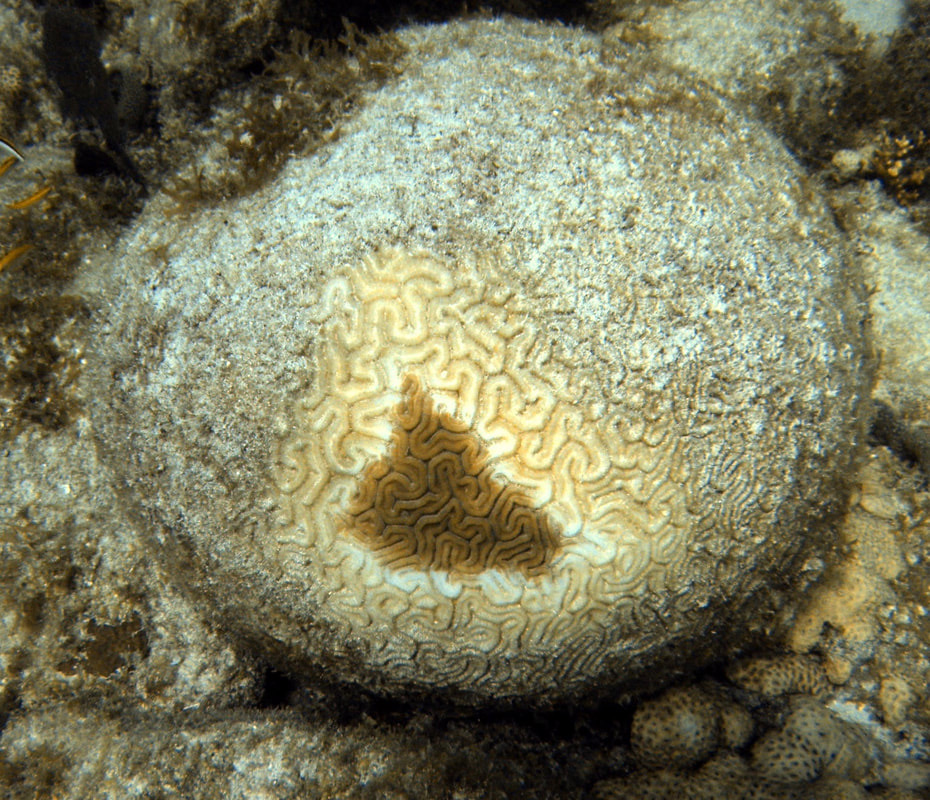
Another coral almost completely dead from stony coral tissue loss disease.
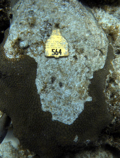
Throughout the Keys, there are tagged coral colonies that are being monitored. The tag has information on it for scuba divers and snorkelers on how to submit photos of these corals! It is a great way for citizen science engagement and community monitoring - I believe AGRRA is the monitoring organization.
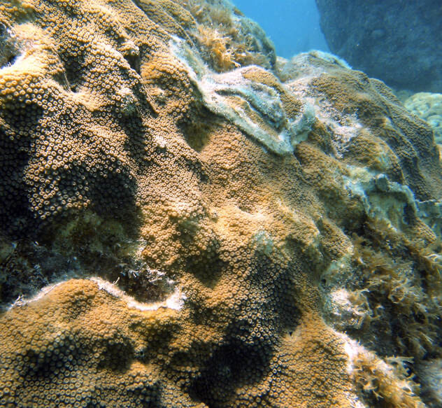
This coral was treated for stony coral tissue loss disease - you can tell by the white, thin paste that was applied to the diseased line. Scientists have found that applying this paste with an antibiotic helps some of these corals by stopping the spread of the disease.
Fun diving with Dr. Grace Klinges
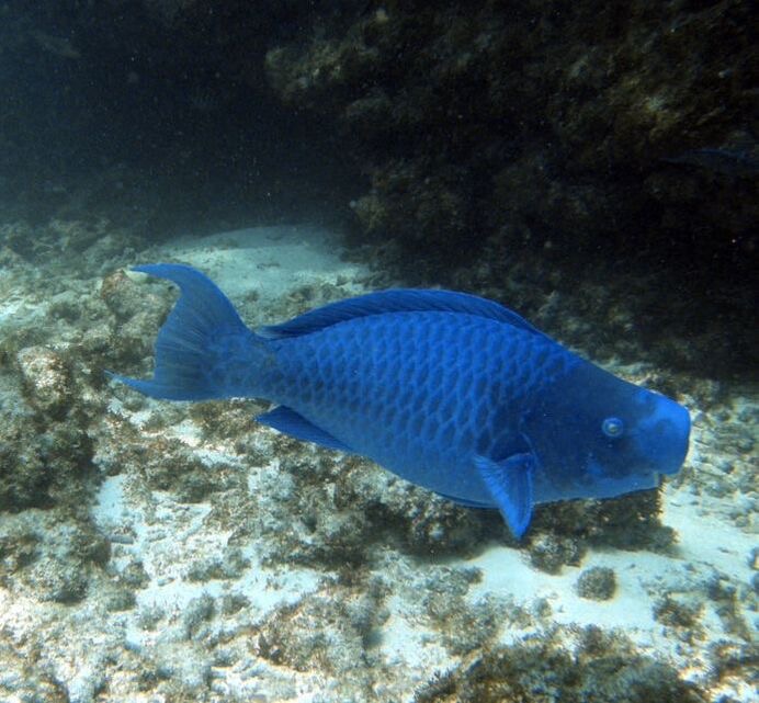
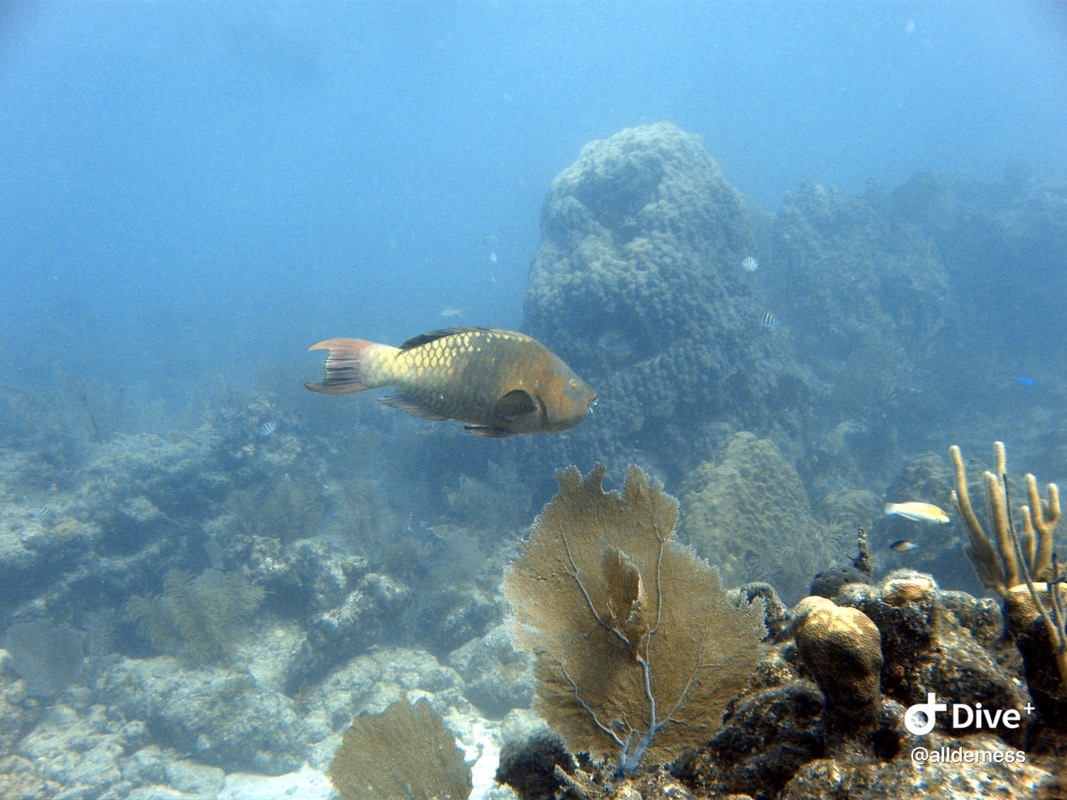
Blue parrotfish - did you know parrotfish chomp on corals in order to eat the algae? Then they digest the coral skeleton and poop out sand!
Acropora palmata outplants
Sarah Hamlyn monitoring coral outplants
Photo by Melissa Heres - fun dive with Rainbow Reef in Key Largo!
Fun dive with JD Reinbott in Looe Key!
A giant Orbicella faveolata. I was shocked!!
JD posing with Acropora palmata
Dendrogyra cylindrus!!!! Super rare coral in the Keys at this point. It was so exciting to see a healthy colony!
Close-up on some Orbicella faveolata
Orbicella faveolata with a neon goby hanging out!
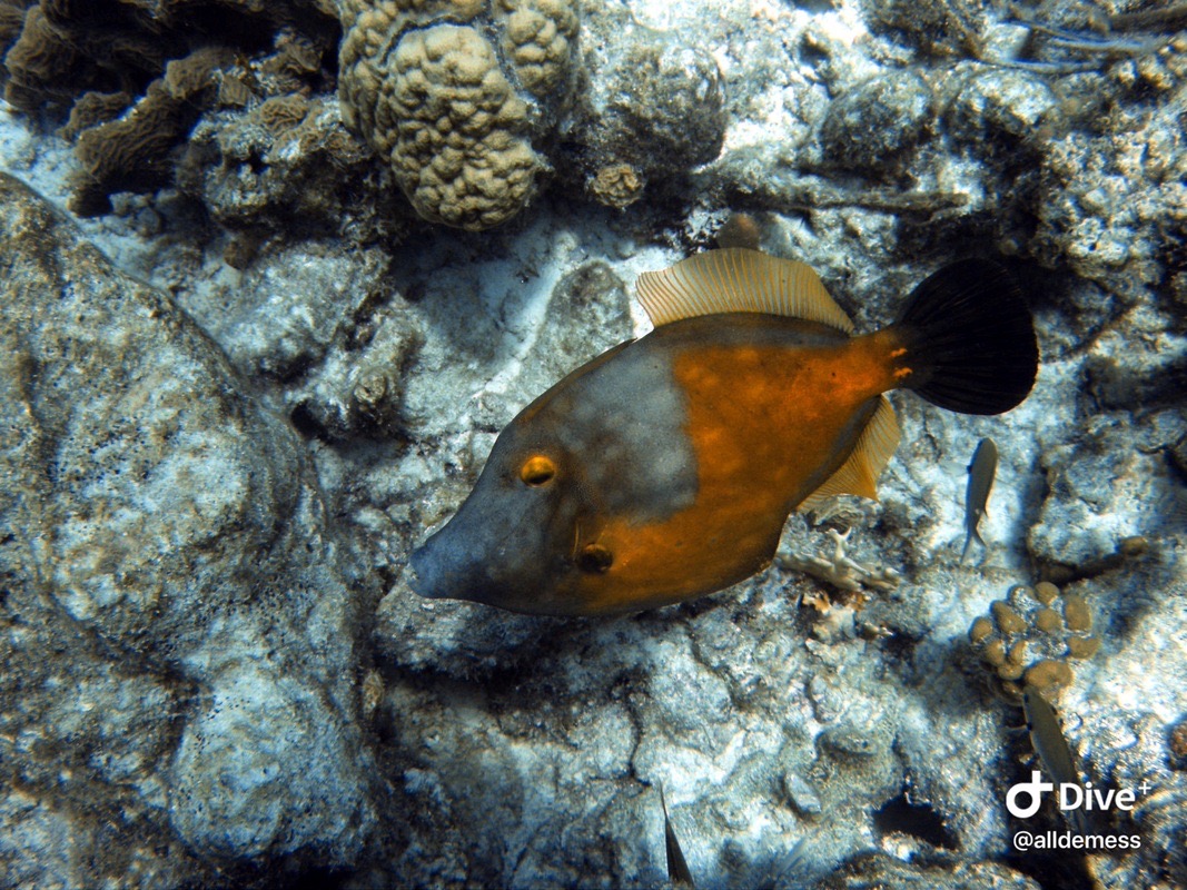
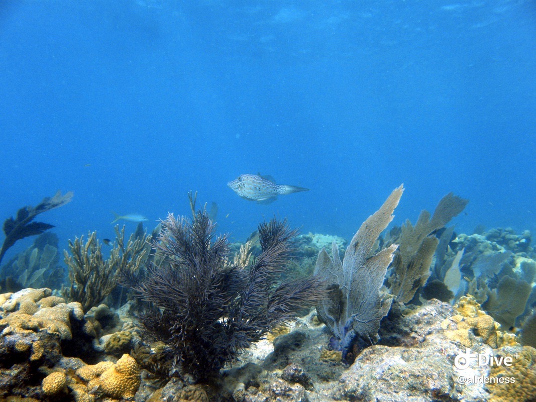
Scrawled filefish zooming by
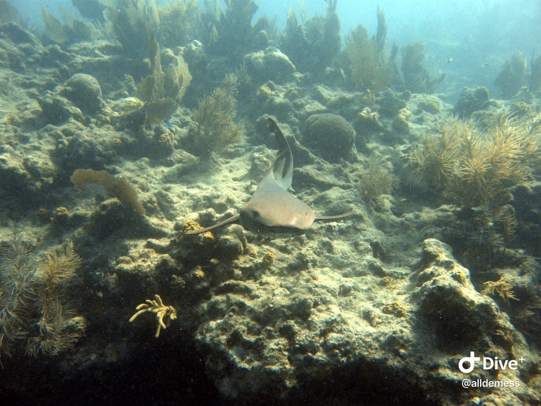
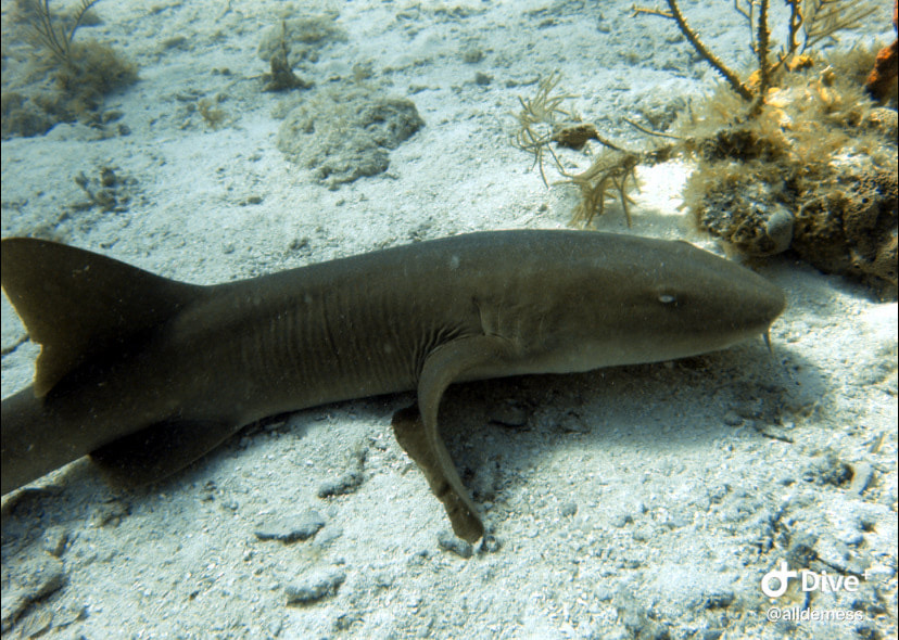
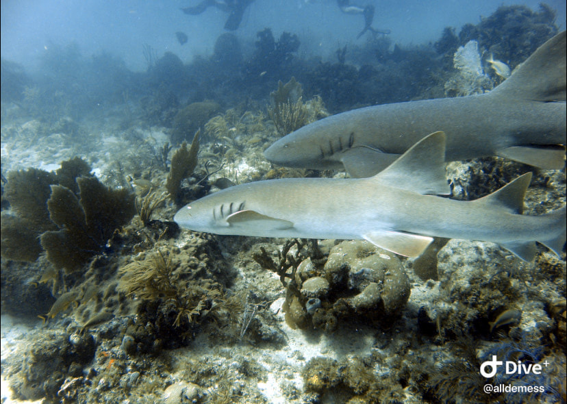
A very friendly nurse shark
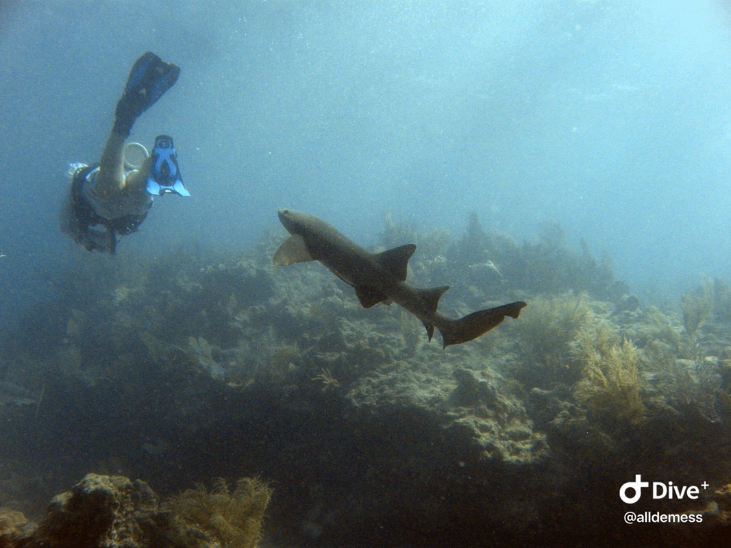
The nurse shark going for JD’s fin!
Acropora palmata and Colpophyllia natans growing together
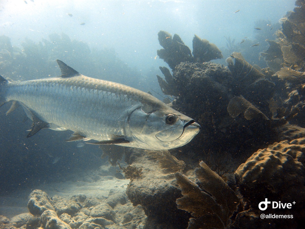
A giant tarpon that spooked me
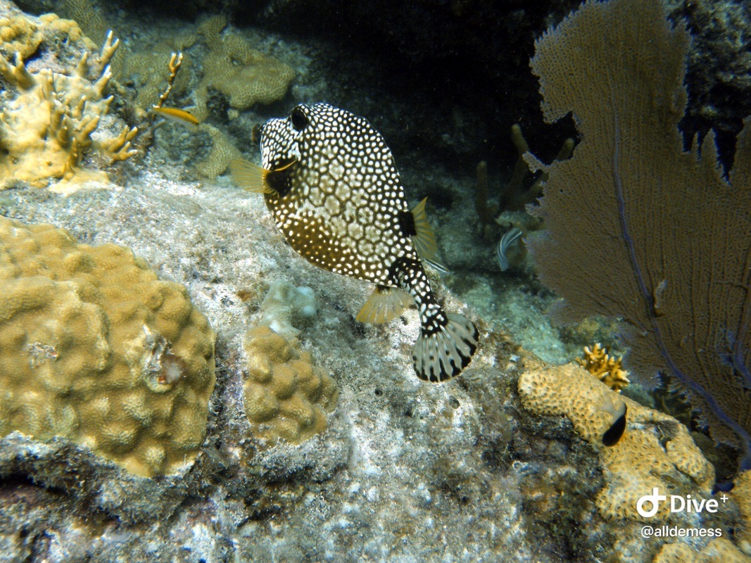
A smooth trunkfish! My favorite fish, so cute
Acropora palmata outplants from Coral Restoration Foundation
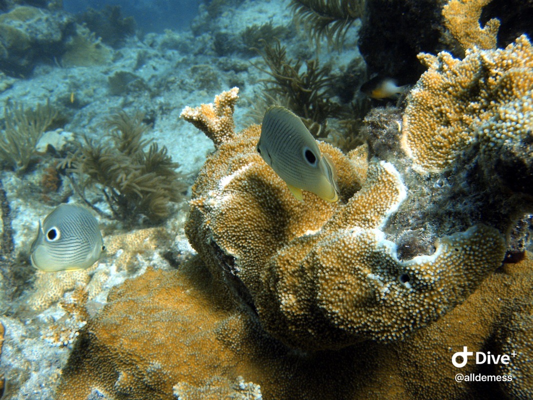
Foureye butterflyfish - they always hang out in pairs!
JD with some healthy Acropora palmata
JD with the sad state of the reef landscape - this is a lot of what you see nowadays.
Close-up of some fluffy Dendrogyra cylindrus
Stony coral tissue loss disease progression - I believe that nail was placed there to mark the line of the disease.
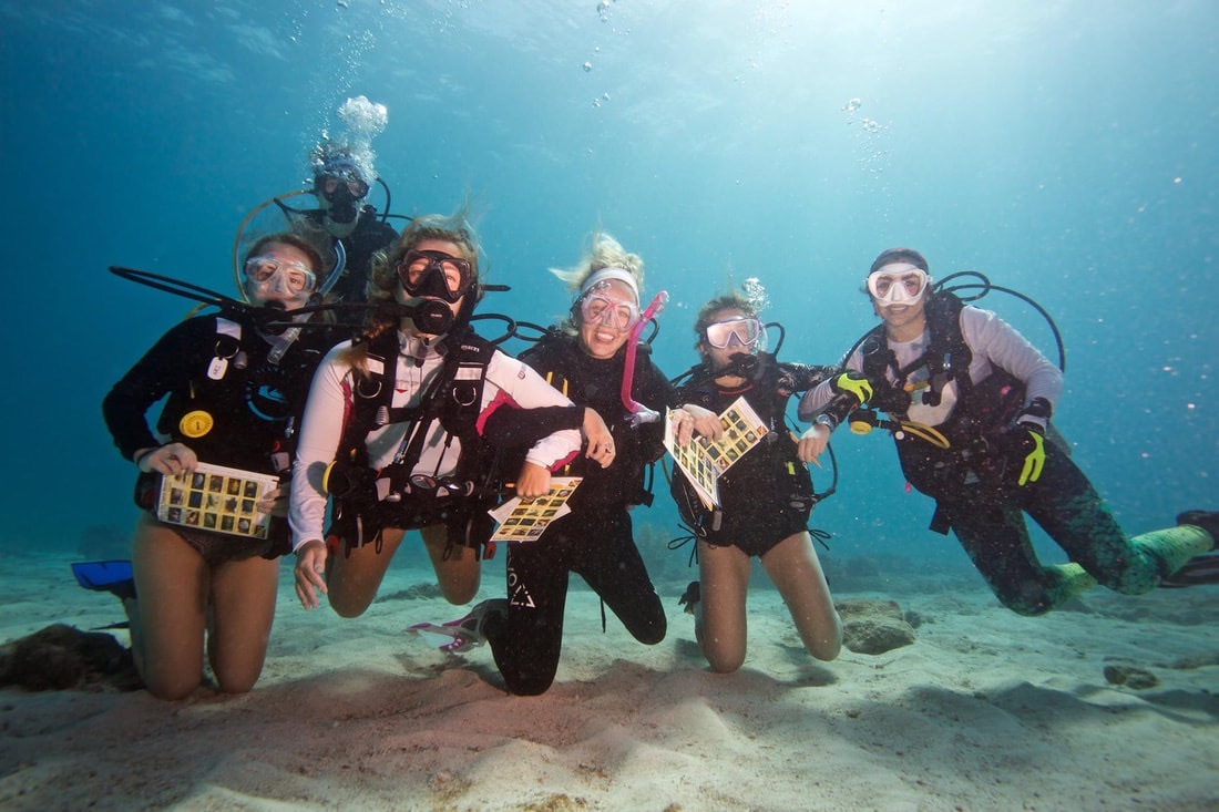
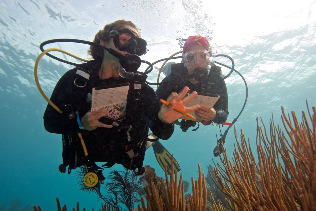
Photo by Carlos Estape - REEF interns on our first fish ID dive! From left to right: Ali Treen, Ellie Place, me, Christy Ascione, Annie Innes-Gold, Sophie Costa
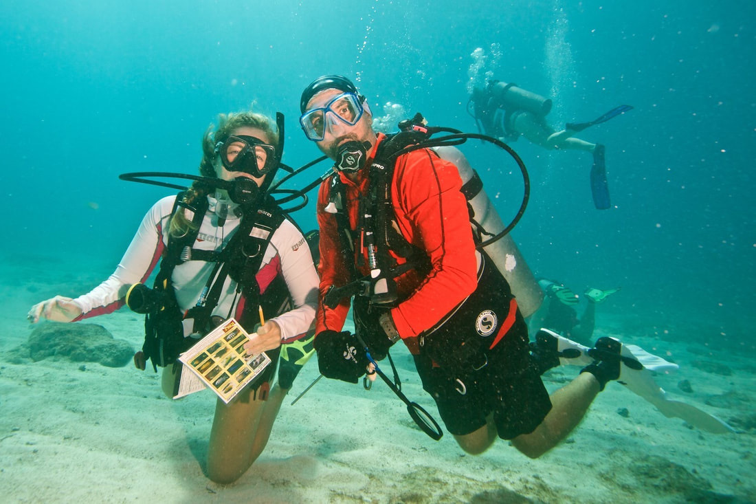
Photo by Allison Estape - myself and Christy looking for some fish
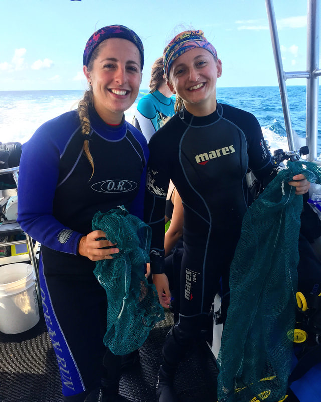
Annie Innes-Gold and I on a clean-up dive against debris with Key Dives shop!
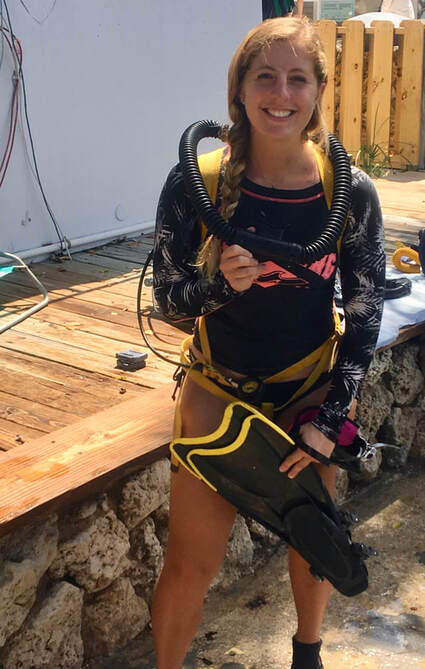
Got to try a double-hose regulator with the History of Diving Museum at Jules’ Underwater Lodge!
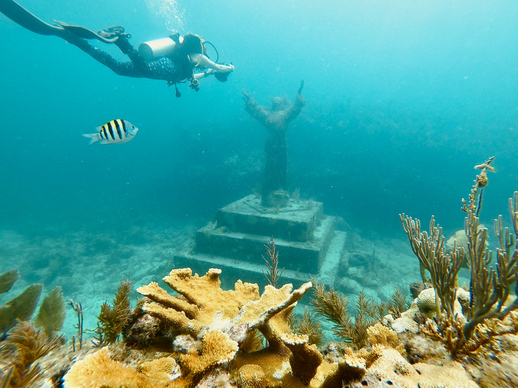
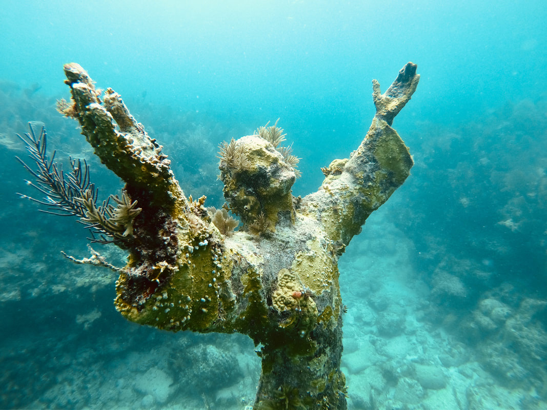
Christ of the Abyss statue, Key Largo
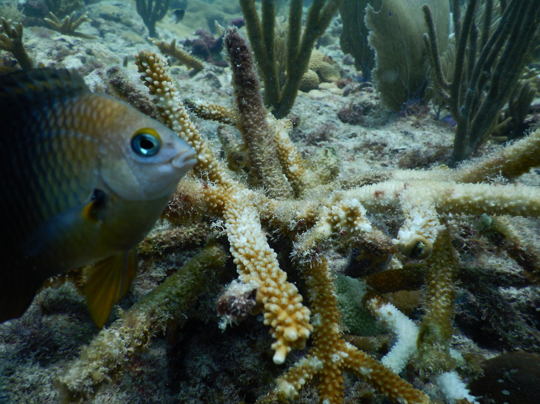
This damselfish was very protective of the staghorn coral colony (which did not look to be in good shape).
bite marks from damselfish
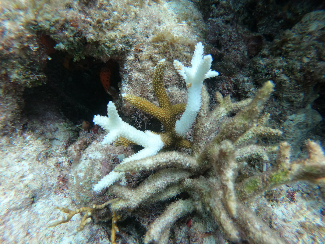
disease on Acropora cervicornis
Montastrea cavernosa (photo by Martine D’Alessandro)
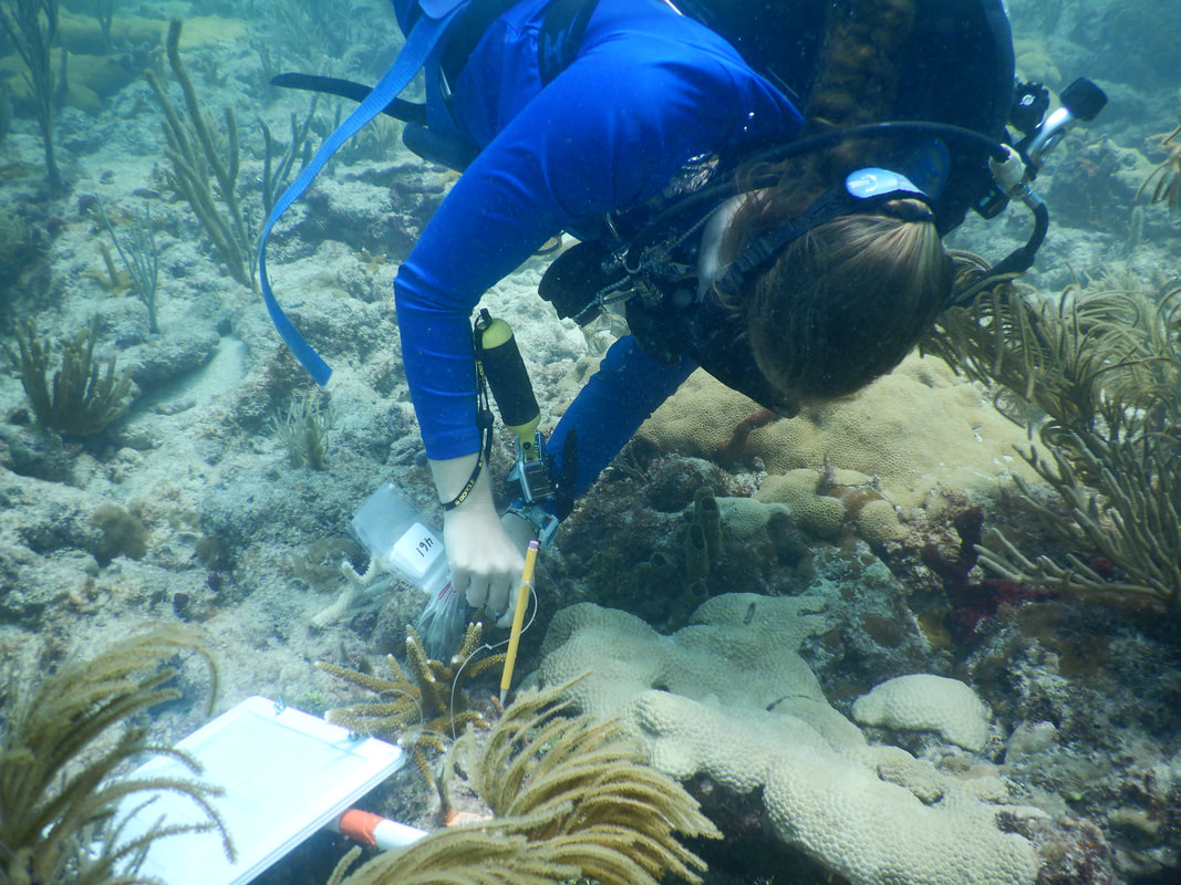
sampling a diseased coral
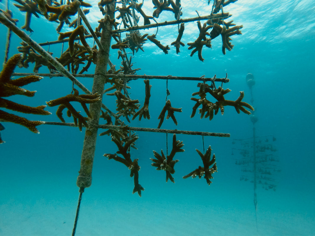
nursery tree (Lirman Lab / Rescue a Reef)
Acropora cervicornis nursery tree
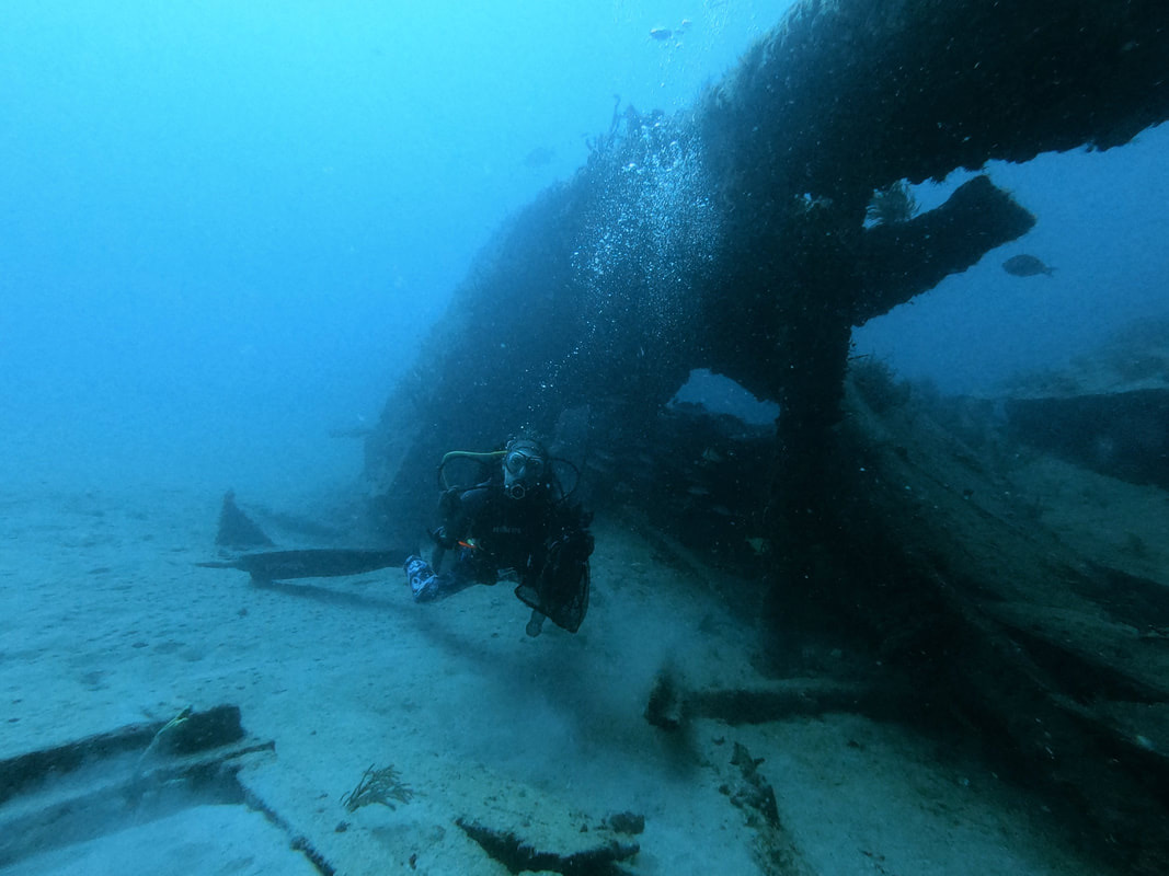
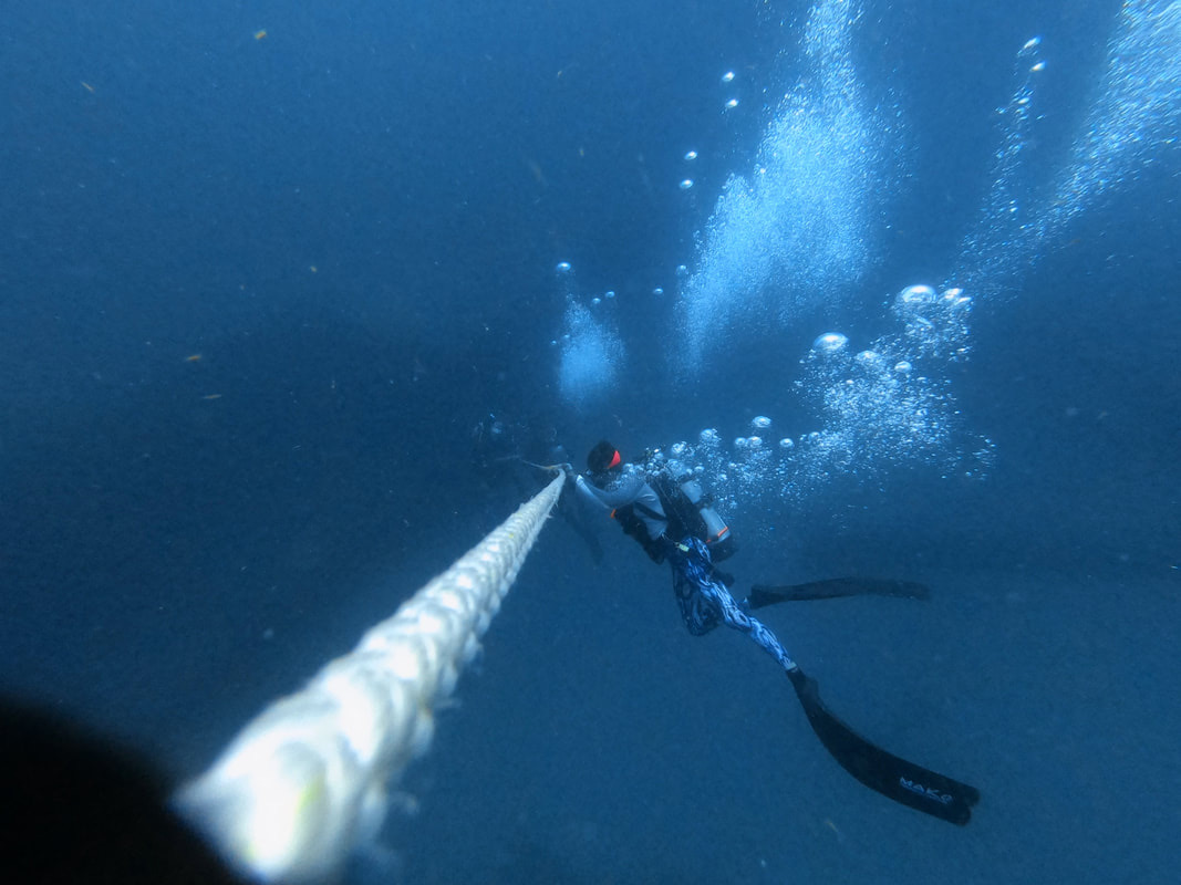
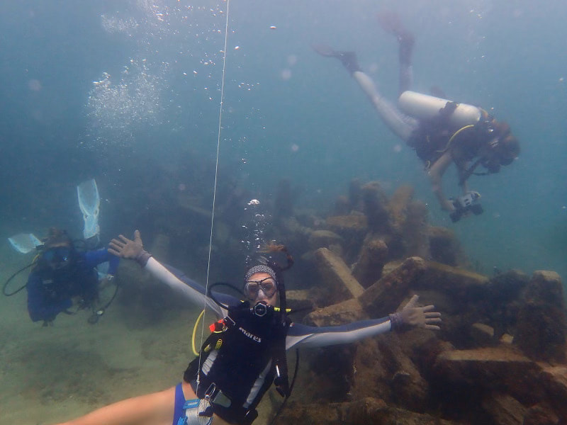
going down the line to a wreck off Fort Lauderdale
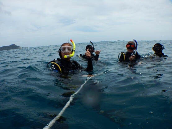
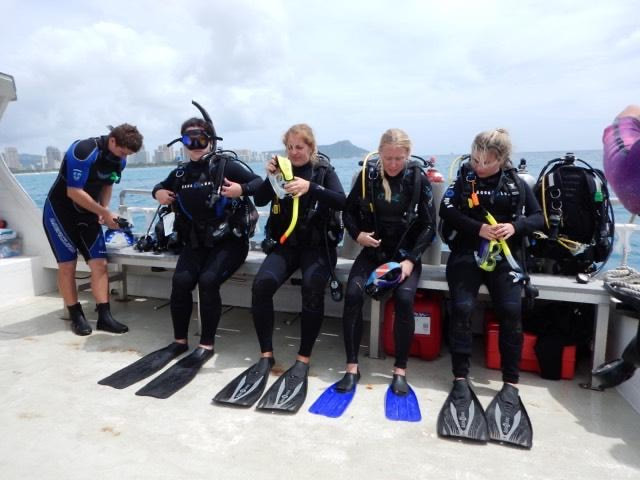
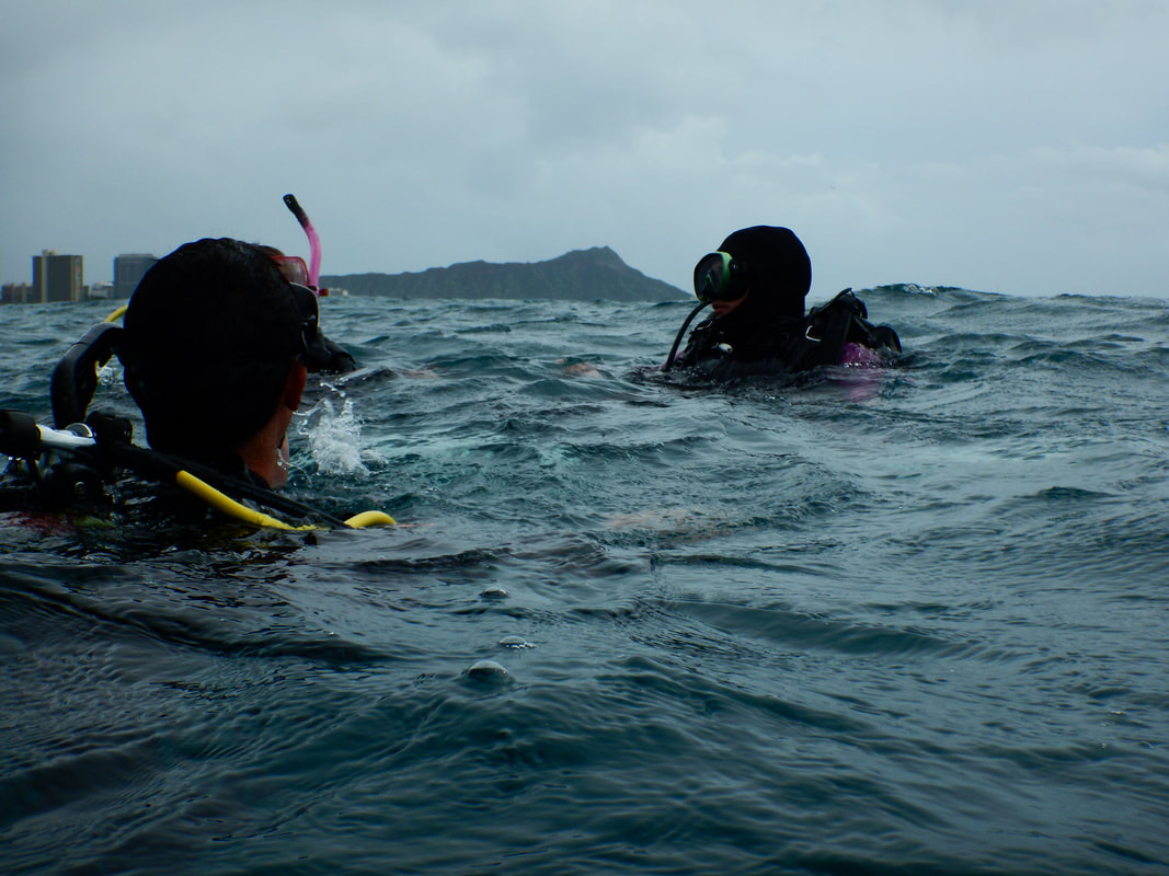
First open water dive in Honolulu (Diamond Head in the background)
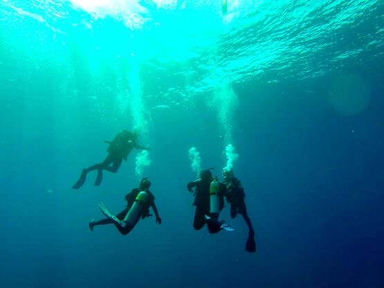
First open water dive in Honolulu
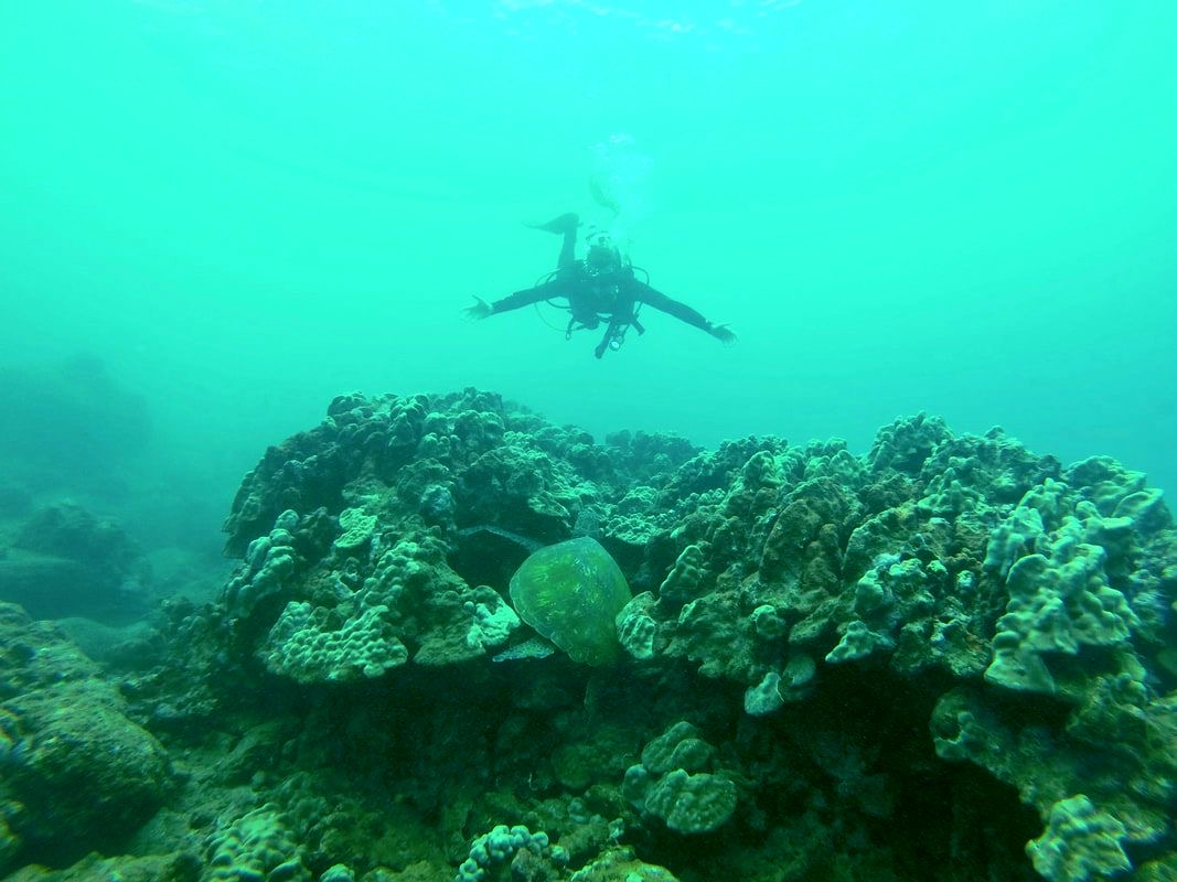
Diving in Kauai (spot the turtle!)
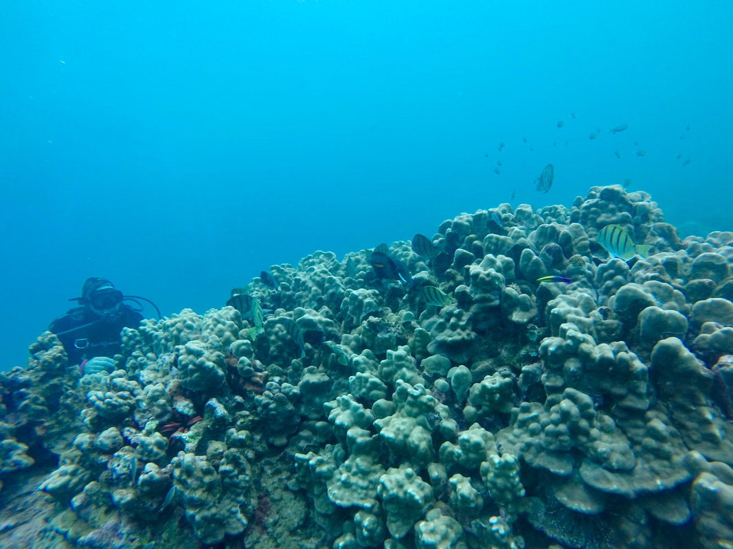
First open water dive in Honolulu
First open water dive in Honolulu